. 2009;4(2):e4309.
doi: 10.1371/journal.pone.0004309. Epub 2009 Feb 3.

Sequential metabolism of 7-dehydrocholesterol to steroidal 5,7-dienes in adrenal glands and its biological implication in the skin

Affiliations

Sequential metabolism of 7-dehydrocholesterol to steroidal 5,7-dienes in adrenal glands and its biological implication in the skin

Andrzej T Slominski et al. PLoS One. 2009.

Abstract

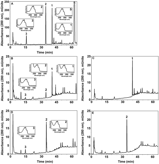
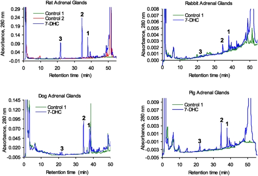
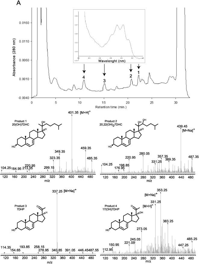
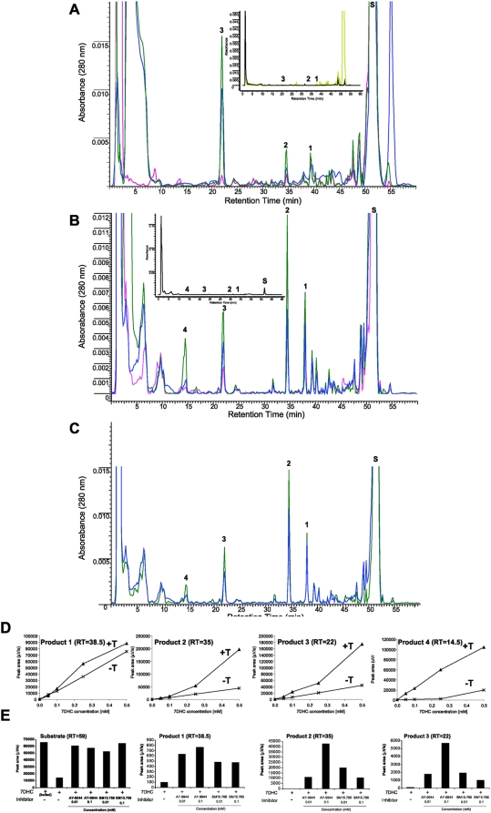
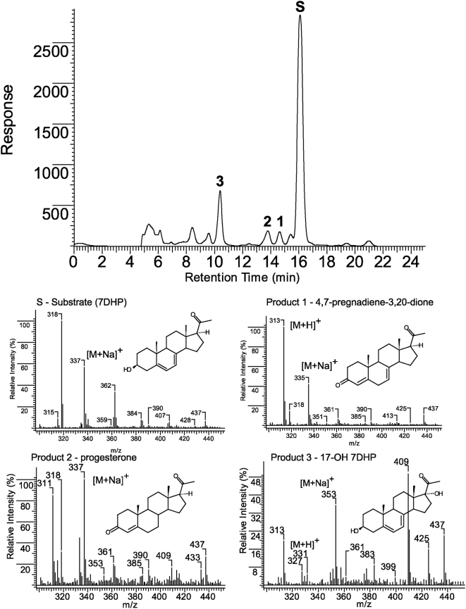
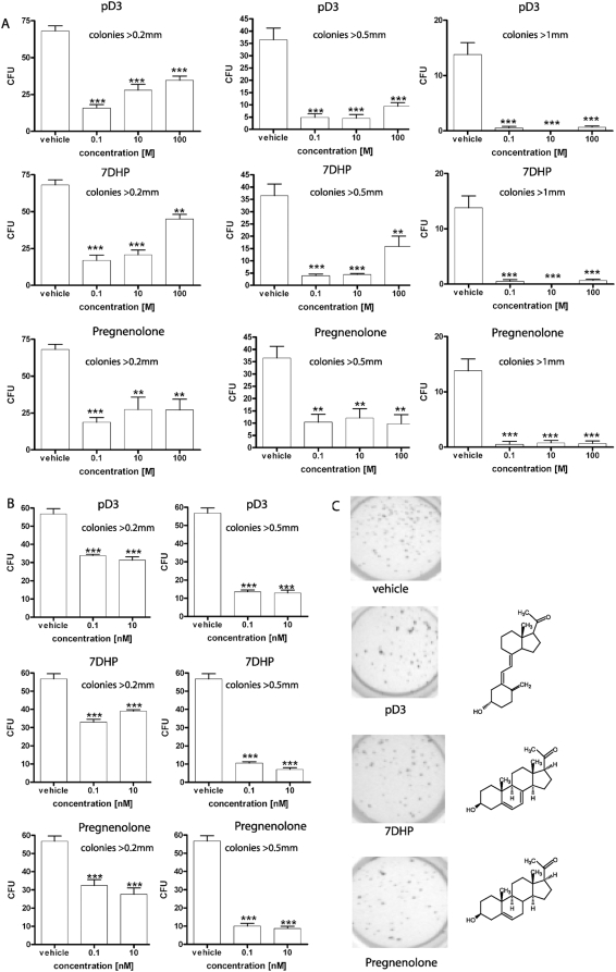
Since P450scc transforms 7-dehydrocholesterol (7DHC) to 7-dehydropregnenolone (7DHP) in vitro, we investigated sequential 7DHC metabolism by adrenal glands ex vivo. There was a rapid, time- and dose-dependent metabolism of 7DHC by adrenals from rats, pigs, rabbits and dogs with production of more polar 5,7-dienes as detected by RP-HPLC. Based on retention time (RT), UV spectra and mass spectrometry, we identified the major products common to all tested species as 7DHP, 22-hydroxy-7DHC and 20,22-dihydroxy-7DHC. The involvement of P450scc in adrenal metabolic transformation was confirmed by the inhibition of this process by DL-aminoglutethimide. The metabolism of 7DHC with subsequent production of 7DHP was stimulated by forscolin indicating involvement of cAMP dependent pathways. Additional minor products of 7DHC metabolism that were more polar than 7DHP were identified as 17-hydroxy-7DHP (in pig adrenals but not those of rats) and as pregna-4,7-diene-3,20-dione (7-dehydroprogesterone). Both products represented the major identifiable products of 7DHP metabolism in adrenal glands. Studies with purified enzymes show that StAR protein likely transports 7DHC to the inner mitochondrial membrane, that 7DHC can compete effectively with cholesterol for the substrate binding site on P450scc and that the catalytic efficiency of 3betaHSD for 7DHP (V(m)/K(m)) is 40% of that for pregnenolone. Skin mitochondria are capable of transforming 7DHC to 7DHP and the 7DHP is metabolized further by skin extracts. Finally, 7DHP, its photoderivative 20-oxopregnacalciferol, and pregnenolone exhibited biological activity in skin cells including inhibition of proliferation of epidermal keratinocytes and melanocytes, and melanoma cells. These findings define a novel steroidogenic pathway: 7DHC-->22(OH)7DHC-->20,22(OH)(2)7DHC-->7DHP, with potential further metabolism of 7DHP mediated by 3betaHSD or CYP17, depending on mammalian species. The 5-7 dienal intermediates of the pathway can be a source of biologically active vitamin D3 derivatives after delivery to or production in the skin, an organ intermittently exposed to solar radiation.


Conflict of interest statement

Competing Interests: The authors have declared that no competing interests exist.

Figures

Figure 1
Figure 1. 7DHC metabolism by rat adrenal glands.
Finely cut fragments of rat adrenal glands were incubated in the presence (B–D) or absence (A) of 7DHC (0.5 mM). Boiled adrenal fragments (C) served as an additional negative control. Panel B shows results from an incubation which included trilostane, a 3βHSD inhibitor. Steroids were extracted with methylene chloride and subjected to LC/MC and UV spectra analyses. Panels A–D identifies 7 main products. Product 3 had an identical retention time to 7DHP standard. Panel E shows UV spectra, MS and the predicted structures of the 7 metabolites.
Figure 2
Figure 2. Precursor-product relationship between 7DHC, hydroxy-7DHC, dihydroxy-7DHC and 7DHP defined by their sequential metabolism by rat adrenal glands.
A. Chromatogram showing metabolism of 7DHC(S) by adrenal glands. The hydroxy-7DHC (1) and dihydroxy-7DHC (2) products were collected from the HPLC and tested as substrates (see B–E). Product 3 was identified as 7-DHP based on RT, mass and UV spectra. B. Incubation of hydroxy-7DHC (1) with adrenal glands generated dihydroxy-7DHC with RT 34.8 min (2) and 7DHP with RT 15.8 min (3). C. Boiled adrenal glands served as a control for B. D. Incubation of dihydroxy-7DHC (2) with adrenal glands produced 7DHP; note the lack of S and product 1. E. Negative control for D (boiled adrenal glands ). Inserts: UV spectra of compounds 1–3.
Figure 3
Figure 3. Metabolism of 7-DHC by adrenal glands of different mammalian species.
Finely cut fragments of rat (A), rabbit (B), dog (C) and pig (D) adrenals were incubated in the presence (blue) or absence (green) of 7DHC (S). Boiled adrenal fragments served as an additional negative control for the rat (red). The three major products common to all animals studied are numbered 1 to 3 on the chromatograms. Product 3 had an identical retention time, mass, and UV spectra to 7DHP standard.
Figure 4
Figure 4. Metabolism of 7DHC by pig adrenal glands.
Fragments of pig adrenal glands were incubated with 0.5 mM 7DHC. Steroids were extracted with methylene chloride and subjected to HPLC and UV spectral analyses. The UV spectrum for all peaks is the same (see arrows). The mass spectra of the collected peaks 1–4 were obtained by direct injection into the MS module of the Bruker Esquire-LC/MS Spectrometer.
Figure 5
Figure 5. Metabolism of 7DHC by adrenal glands in the presence of DL-aminoglutethimide (AGT), trilostane, forscolin and Δ7-reductase inhibitors.
A. For rat adrenals transformation of 7DHC (S) to 7DHP (3) was dramatically inhibited by AGT (red) compared to the incubation with 7DHC alone (blue), while accumulation of 22(OH)7DHC (1), 20,22(OH)27DHC (2) and 7DHP (3) was enhanced by trilostane (green). Control incubations performed with boiled adrenal fragments are shown in the inset. Incubations were carried out for 18 h. B. For pig adrenals AGT also inhibited 7DHC metabolism (red) compared to the incubation with 7DHC alone (blue), while trilostane (green) enhanced accumulation of 22(OH)7DHC (1), 20,22(OH)27DHC (2), 7DHP (3) and 17(OH)7DHP (4). Adrenal fragments were incubated for 6 h and controls (inset) were incubated without the substrate. C. For pig adrenals 0.1 mM forscolin (green) stimulated 7DHC metabolism. The labeling and the experimental conditions are the same as in B. D. Fragments of pig adrenal glands were incubated with increasing doses of 7DHC in the presence (T+) or absence (T−) of 0.1 mM trilostane. The chromatograms were processed as described above. Trilostane (T) enhanced accumulation of 22(OH)7DHC (1), 20,22(OH)27DHC (2), 7DHP (3) and 17(OH)7DHP (4) as a function of 7DHC concentration. E. Fragments of rat adrenal glands were incubated with 7DHC (0.5 mM) in presence of AY-9944 or BM15.766 (10 µM or 100 µM) and analyzed by RP-HPLC (see A for peak identification). The chromatograms were processed using ACDLabs software and relative areas for peaks identified above were calculated. Δ7-reductase inhibitors enhanced production of 7DHC metabolites.
Figure 6
Figure 6. Time-dependent metabolism of 7DHP is modified by trilostane (T).
Fragments of the pig adrenal glands were incubated with 7DHP (S; 0.5 mM) in the absence (A) or presence (B) of trilostane (0.1 mM) and analyzed by RP-HPLC. Boiled adrenal fragments were used as a negative control (blue). The major products of 7DHP metabolism affected by trilostane are marked by numbers. Time-dependent changes in the relative concentration of the identified products in presence (T+) or absence (T-) of trilostane were calculated using ACDLabs software and are presented in C.
Figure 7
Figure 7. Trilostane (T) inhibits and modifies 7DHP (S) metabolism by rat adrenal glands.
A. Fragments of rat adrenal glands were incubated with 7DHP (0.5 mM) in the absence (pink) or presence (gray) of trilostane (0.1 mM) and samples analyzed by RP-HPLC. Boiled adrenal fragments (green) or incubations without the substrate (blue) were used as negative controls. B. The relative concentrations of the identified products affected by trilostane (T). The insert shows inhibition of 7DHP consumption by trilostane. Control marked by blue represents or incubations without the substrate.
Figure 8
Figure 8. LC/MS identification of the products of 7DHP metabolism by pig adrenals.
Collected fraction from RT-HPLC experiment were analyzed using the Bruker Esquire-LC/MS Spectrometer as described in Materials and Methods. Numbers correspond to the products identified in Figs 6 and 7.
Figure 9
Figure 9. Proposed pathways for the in vivo metabolism of 7DHC in the mammalian adrenal gland.
Figure 10
Figure 10. Stimulation of 7DHC transfer between membranes and the inhibition of the side chain cleavage of [cholesterol by 7DHC.
A. To study 7DHC transfer between membranes by N-62 StAR protein cholesterol or 7DHC was placed in donor vesicles at a molar ratio to phospholipid of 0.2. The transfer of the sterol to acceptor vesicles at 35°C was measured in the presence or absence of 5 µM N-62 StAR protein. B. Inhibition of the side chain cleavage of [4-14C]cholesterol by 7DHC was determined with substrate and cytochrome P450scc incorporated into phospholipid (PL) vesicles prepared from dioleoyl phosphatidylcholine containing ardiolipin. As indicated in the figure, 7DHC was included in the membrane at a molar ratio to phospholipid of 0.2. The rate of side chain cleavage of [4-14C]cholesterol was determined from the amount of [4-14C]pregnenolone formed.
Figure 11
Figure 11. The effect of 7DHP, pregnacholecalciferol (pD3) and pregnenolone on normal immortalized epidermal HaCaT keratinocytes and PIG1 melanocytes.
A and B. Inhibition of HaCaT cell proliferation as measured by [3H]-thymidine incorporation (A) and MTT (B) assays after 48 h and 72 h of treatment, respectively. C. Inhibition of NFκB activity in HaCaT keratinocytes 24 h after transfection with luciferase construct NFκB-Luc. The cells were treated for 24 h with 100 nM vitamin D3 (vD3), 1,25(OH)2D3, 7DHP, pD3, pregnenolone or ethanol vehicle (control) and luciferase activity was measured in cell extracts. D and E. Inhibition of cell growth as measured by sulforhodamine b assay (protein content) after 72 h of treatment of non-pigmented (D) and pigmented (E) PIG1 human melanocytes. Inserts show the color of representative cell pellets. Significant differences versus ethanol-treated control cells were defined as *p<0.05, **p<0.01, ***p<0.001.
Figure 12
Figure 12. 7DHP, 20-oxopregnacalciferol (pD3) and pregnenolone inhibit growth of melanoma cells in soft agar.
A: hamster AbC1 melanoma; B: human SKMEL-188 melanoma; C: visualization of SKMEL-188 colonies by MTT reagent. Decreased number and size of colonies was observed in both cell lines. Significant differences versus ethanol-treated cells were defined as *p<0.05, **p<0.01, ***p<0.001.
Figure 13
Figure 13. Metabolism of 7DHC and 7DHP by rat skin.
A. To study the skin capability to transform 7DHC to 7DHP isolated mitochondria were incubated with 7DHC (0.5 mM) in the presence (experimental) or absence (control) of isocitrate and NADPH. 7DHP: product with the same retention time and UV spectra (inset) as 7DHP standard. B. Incubation of crude skin extract with 7DHP substrate (RT 16.7 min) in the presence of isocitrate and NADPH (experimental) resulted in appearance of a 5,7-diene product (N) with RT 11.9 min, which was absent in control reaction mixture (isocitrate and NADPH were not added) (control).

References

    1. Payne AH, Hales DB. Overview of steroidogenic enzymes in the pathway from cholesterol to active steroid hormones. Endocr Rev. 2004;25:947–70. - PubMed
    1. Tuckey RC. Progesterone synthesis by the human placenta. Placenta. 2005;26:273–81. - PubMed
    1. Burstein S, Gut M. Intermediates in the conversion of cholesterol to pregnenolone: kinetics and mechanism. Steroids. 1976;28:115–31. - PubMed
    1. Hume R, Kelly RW, Taylor PL, Boyd GS. The catalytic cycle of cytochrome P-450scc and intermediates in the conversion of cholesterol to pregnenolone. Eur J Biochem. 1984;140:583–91. - PubMed
    1. Lambeth JD, Seybert DW, Lancaster JR, Jr, Salerno JC, Kamin H. Steroidogenic electron transport in adrenal cortex mitochondria. Mol Cell Biochem. 1982;45:13–31. - PubMed

Publication types

MeSH terms

LinkOut - more resources