Comparative Study
. 2009 May;467(5):1243-9.
doi: 10.1007/s11999-008-0699-0. Epub 2009 Feb 4.

CT study on the effect of different treatment protocols for clubfoot pathology

Affiliations
Comparative Study

CT study on the effect of different treatment protocols for clubfoot pathology

Pasquale Farsetti et al. Clin Orthop Relat Res. 2009 May.

Abstract

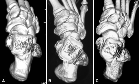
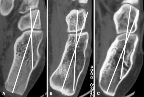
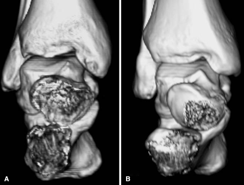
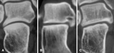
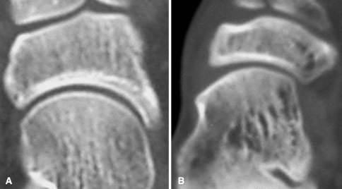
In congenital clubfoot, residual deformities are not well-documented and they may change depending on different treatments. To identify the treatment that provides better outcome at maturity, we studied the computed tomography of two cohorts of patients affected with congenital clubfoot who were treated using two distinct protocols. Forty-seven clubfeet were treated according to the traditional protocol of our hospital and 61 were treated according to the Ponseti technique. The normal feet of the unilateral deformities served as controls. All patients were followed to skeletal maturity. The ankle torsion angle and the declination angle of the neck of the talus were higher than normal but different only in patients treated with the traditional method. The calcaneocuboid angle was lower but only in patients treated with the Ponseti method. The shape of the talar joints was altered in many feet regardless of protocol. The CT images suggest the modifications of the torsion angle of the ankle, the declination angle of the neck of the talus, and the calcaneocuboid angle at maturity are related to the treatment protocol followed. The Ponseti manipulative technique provided better anatomical results in comparison to our traditional technique.


Figures

Fig. 1A–C
Fig. 1A–C
Three-dimensional computed tomography scan reconstruction of the foot in the transverse plane at the level of the ankle mortise in (A) a normal foot, (B) a clubfoot of the first series, and (C) a clubfoot of the second series. The torsion angle of the ankle measured 19° in the normal foot, 30.5° in the clubfoot of the first series, and 26.5° in the clubfoot of the second series. The declination angle of the neck of the talus measured 18° in the normal foot, 31° in the first series clubfoot, and 23.5 in the second series clubfoot. A residual varus deformity of the calcaneus is present in the clubfoot of the first series. The navicular tuberosity is very close to the medial malleolus in the clubfoot of the second series, but the cuneiforms and the cuboid are shifted laterally.
Fig. 2A–C
Fig. 2A–C
Computed tomography image of the calcaneocuboid joint (A) in a normal foot, (B) in a clubfoot of the first series, in which the joint appears inverted, and (C) in a clubfoot of the second series, in which it appears flat. The calcaneocuboid angle measured 19.5° in the normal foot, 15.5° in the clubfoot of the first series, and 13° in the clubfoot of the second series.
Fig. 3A–B
Fig. 3A–B
Three-dimensional computed tomography scan reconstruction of the hindfoot in the coronal plane in (A) a clubfoot of the first series and (B) a clubfoot of the second series. Talus and calcaneus are coincident in the clubfoot of the first series and divergent in the clubfoot of the second series.
Fig. 4A–C
Fig. 4A–C
Computed tomography image of the subtalar joint at the level of the posterior articulation in (A) a normal foot, (B) a clubfoot of the second series, in which it appears flat, and (C) a clubfoot of the first series, in which the joint appears slanted.
Fig. 5A–B
Fig. 5A–B
Computed tomography image of the talonavicular joint in (A) a normal foot and (B) in a clubfoot of the second series, in which it is medially subluxated.

References

    1. Codivilla A. Sulla cura del piede equino varo congenito: nuovo metodo di cura cruenta [Congenital talipes equinovarus: a new method of surgical treatment] Arch Chir Orthop. 1906;23:245–258.
    1. Farsetti P, Caterini R, Mancini F, Potenza V, Ippolito E. Anterior tibial tendon transfer in relapsing congenital clubfoot. J. Pediatr. Orthop. 2006;26:83–90. doi: 10.1097/01.bpo.0000188999.21427.fb. - DOI - PubMed
    1. Howard CB, Benson MKD. Clubfoot: its pathological anatomy. J. Pediatr. Orthop. 1993;13:654–659. - PubMed
    1. Ippolito E, Farsetti P, Caterini R, Tudisco C. Long-term comparative results in patients with congenital clubfoot treated with two different protocols. J. Bone Joint Surg. Am. 2003;85:1286–1294. - PubMed
    1. Ippolito E, Fraracci L, Caterini R, Di Mario M, Farsetti P. A radiographic comparative study of two series of skeletally mature clubfeet treated by two different protocols. Skeletal Radiol. 2003;32:446–453. doi: 10.1007/s00256-003-0620-6. - DOI - PubMed

Publication types