Review
. 2009 Jan;16(1):58-83.
doi: 10.1080/10739680802651477.

Platelet adhesion under flow

Affiliations
Review

Platelet adhesion under flow

Zaverio M Ruggeri. Microcirculation. 2009 Jan.

Abstract

Platelet-adhesive mechanisms play a well-defined role in hemostasis and thrombosis, but evidence continues to emerge for a relevant contribution to other pathophysiological processes, including inflammation, immune-mediated responses to microbial and viral pathogens, and cancer metastasis. Hemostasis and thrombosis are related aspects of the response to vascular injury, but the former protects from bleeding after trauma, while the latter is a disease mechanism. In either situation, adhesive interactions mediated by specific membrane receptors support the initial attachment of single platelets to cellular and extracellular matrix constituents of the vessel wall and tissues. In the subsequent steps of thrombus growth and stabilization, adhesive interactions mediate platelet-to-platelet cohesion (i.e., aggregation) and anchoring to the fibrin clot. A key functional aspect of platelets is their ability to circulate in a quiescent state surveying the integrity of the inner vascular surface, coupled to a prompt reaction wherever alterations are detected. In many respects, therefore, platelet adhesion to vascular wall structures, to one another, or to other blood cells are facets of the same fundamental biological process. The adaptation of platelet-adhesive functions to the effects of blood flow is the main focus of this review.


Figures

Figure 1
Figure 1. Schematic representation of blood flow in a vessel
Normal endothelial cells are non reactive for platelets, but exposed subendothelial structures induce rapid platelet adhesion and aggregation. Blood flow in a cylindrical vessel can be visualized as a series of fluid layers (laminae) moving at different velocity. The laminae near the center of the vessel have greater velocity than those near the wall (depicted by arrows of different length). The corresponding velocity profile (solid line) is more blunted than the parabolic profile expected with a homogeneous suspension (dotted line) because of cell depletion in the boundary layer near the wall. The shear rate is the rate of change of velocity with respect to distance measured perpendicularly to the direction of flow. The negative sign indicates that the gradient is defined from the center (where velocity is maximal) to the wall (where velocity is minimal).
Figure 2
Figure 2. Platelet count and surface adhesion
These images are derived from a real time experiment recorded at the video rate of 30 frames per second. Whole blood containing the α-thrombin inhibitor D-phenylalanyl-L-prolyl-L-arginine chloromethyl ketone dihydrochloride (PPACK) as the anticoagulant was perfused over surfaces coated with immobilized VWF at the wall shear rate of 1,500 s−1. Each fluorescent (white) particle is a single platelet tethered to the surface. The platelet count in the perfused blood was decreased by sedimenting the cells at low g force, removing the platelet rich plasma layer and replacing it with the same volume of homologous platelet poor plasma. Each image represents a single frame recorded one minute after initiating blood perfusion. Typically, surface coverage was maximal within 10 seconds from the beginning of flow. Note the decrease in surface coverage only at the lowest platelet count tested.
Figure 3
Figure 3. Role of GP Ibα in platelet aggregation during thrombus growth
Blood containing PPACK as the anticoagulant was perfused over collagen type I fibers for 100 seconds at the wall shear rate of 1500 s−1. At this point, the height of thrombi was measured from confocal (z) sections, as shown in the stacks labeled A, while the perfusion continued for an additional 740 seconds with blood containing either buffer or function-blocking monoclonal antibodies directed against GP Ibα or αIIb-β3, or both, as indicated. The flow rate was decreased such that the calculated shear rate was 300 s−1 at the collagen coated surface but >1000 s−1 at the surface of thrombi with a height >20 mμ. After a total perfusion time of 840 seconds, thrombus height was measured by confocal sections, as shown in the stacks labeled B. The results demonstrate that the adhesive function of GP Ibα is as necessary as that of αIIbβ3 to sustain platelet aggregation at the edge of a growing thrombus. Modified from Ruggeri ZM et al [151] and reprinted with permission.
Figure 4
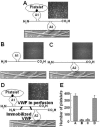
Figure 4. Functional self-association of VWF multimers
A washed blood cells suspensions devoid of plasma proteins and containing EDTA to block integrin function and prostaglandin E1 to block platelet activation was perfused over immobilized collagen type I fibers at the wall shear rate of 1500 s−1. (A) Control experiment with normal multimeric VWF added to the cell suspension. The A3 domain mediates VWF binding to collagen, and the A1 domain interacts with platelet GP Ibα. Tethered platelets are seen rolling on the surface, which is represented by an electron micrograph of collagen fibrils. (B, C) Experiments performed after adding to the cell suspension, respectively, recombinant VWF devoid of the A3 domain (ΔA3-VWF), which cannot bind to collagen, or devoid of the A1 domain (ΔA1-VWF), which binds to collagen but cannot interact with platelet GP Ibα. In either case, no platelets are seen tethered to the surface. (D) Collagen was pre-coated with ΔA1-VWF multimers, which cannot initiate platelet tethering, and then exposed to the blood cell suspension containing ΔA3-VWF. Although the latter cannot bind directly to collagen (see b), it could compensate for the lack of A1 domain in the surface-bound VWF and restore platelet tethering. The association of VWF multimers with one another can explain this result; the two-sided arrows between multimers indicate that the association is reversible. The images are single frames from a real time recording representing an area of 65,536 μm2. The bar graph (E) shows the number of platelets tethered to the surface under the different experimental conditions described above (mean ± SEM of two separate experiments). Modified from Savage B et al [128] and reprinted with permission.
Figure 5
Figure 5. Activation-independent platelet adhesion and aggregation at the interface of immobilized and soluble VWF
(A). Blood containing 93 μM PPACK as anticoagulant, the fluorescent dye mepacrine (10 μM), prostaglandin (PG) E1 (10 μM) to inhibit platelet activation, and EDTA (5 mM) to prevent ligand binding to integrins, was perfused over immobilized VWF (20 μg/ml coating concentration). The white line delimits VWF-coated (to the left) from uncoated glass. Single platelets adhere when the shear rate is 3,000 s−1 (top); rolling aggregates (some identified by arrowheads) form at 20,000 s−1 (bottom). (B). Perfusion over immobilized VWF of washed blood cells suspended in buffer (20 mM Hepes, 150 mM NaCl, pH 7.4). In the absence of soluble VWF, single platelets adhere when the shear rate is 3,000 s−1 (upper left), and fewer single platelets adhere at 24,000 s−1 (upper right). After adding soluble VWF (20 μg/ml), single platelets adhere at 3,000 s−1 (lower left; an arrow points to a single platelet shown for reference), but aggregates form at 24,000 s−1 (lower right; arrows point to a rolling aggregate and an inset highlights a stretched aggregate during stationary adhesion). (C). Perfusion over immobilized VWF of washed blood cells with added soluble VWF and anti-VWF A1 domain monoclonal antibody (NMC-4, 20 μg/ml).133 No platelet adhesion is detected. Modified from Ruggeri ZM et al [169] and reprinted with permission.
Figure 6
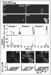
Figure 6. Real time analysis of [Ca++]i during platelet translocation and aggregate formation on immobilized VWF
Platelets loaded with fluo-3 AM (2×10⁷/ml) were suspended with washed erythrocytes in homologous plasma and perfused over immobilized VWF for 3 min at the shear rate of 1500 s−1. The sequence of images at the top shows an example of aggregate formation. At 0 s, platelet 1 appears in the optical field; at 10 s, it has moved in the direction of flow by approximately 20 μm; at 20 s, it has moved by an additional few μm; at 30 s, it is in the same position, and two new platelets (2 and 3) are attached in close proximity forming a small aggregate. The diagrams in the middle show [Ca++]i and instant velocity of platelets 1, 2 and 3. The translocation of platelet 1 occurs mostly during a few seconds of relatively rapid movement, coincident with the appearance of transient [Ca++]i peaks (α/β); a higher and longer lasting increase in [Ca++]i (γ) develops while the platelet is stationary. Cytosolic Ca++ oscillations appear also when platelets 2 and 3 arrest on the surface, without a clear sequence from α/β to γ. The images at the bottom, captured between 60 and 63 s after the appearance of platelet 1 in the field, show the long lasting synchronous increase of [Ca++]i in platelets forming a large aggregate. The 3D diagrams below each image show the measurement of [Ca++]i in all the platelets in the field. Modified from Mazzucato M et al [190]and reprinted with permission.

References

    1. Schulze H, Shivdasani RA. Mechanisms of thrombopoiesis. J Thromb Haemost. 2005;3:1717–1724. - PubMed
    1. Junt T, Schulze H, Chen Z, Massberg S, Goerge T, Krueger A, Wagner DD, Graf T, Italiano JE, Jr., Shivdasani RA, von Andrian UH. Dynamic visualization of thrombopoiesis within bone marrow. Science. 2007;317:1767–1770. - PubMed
    1. Weyrich AS, Lindemann S, Tolley ND, Kraiss LW, Dixon DA, Mahoney TM, Prescott SP, McIntyre TM, Zimmerman GA. Change in protein phenotype without a nucleus: Translational control in platelets. Semin Thromb Hemost. 2004;30:491–498. - PubMed
    1. Weiss HJ. Platelet physiology and abnormalities of platelet function (first of two parts) New Engl J Med. 1975;293:531–541. - PubMed
    1. Weiss HJ. Platelet physiology and abnormalities of platelet function (second of two parts) New Engl J Med. 1975;293:580–588. - PubMed

Publication types