. 2009 Aug 15;57(11):1192-203.
doi: 10.1002/glia.20841.

Human bone marrow-derived mesenchymal stem cells induce Th2-polarized immune response and promote endogenous repair in animal models of multiple sclerosis

Affiliations

Human bone marrow-derived mesenchymal stem cells induce Th2-polarized immune response and promote endogenous repair in animal models of multiple sclerosis

Lianhua Bai et al. Glia. .

Abstract

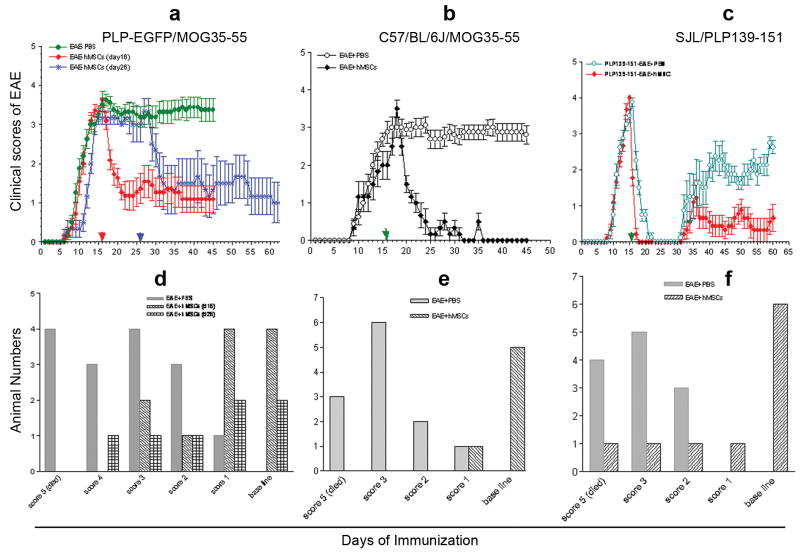
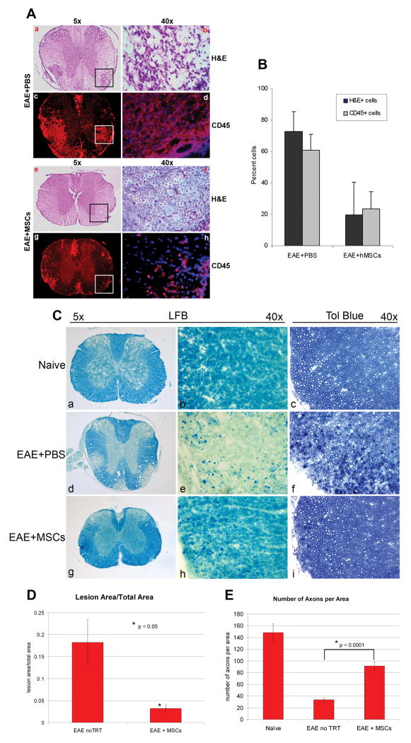
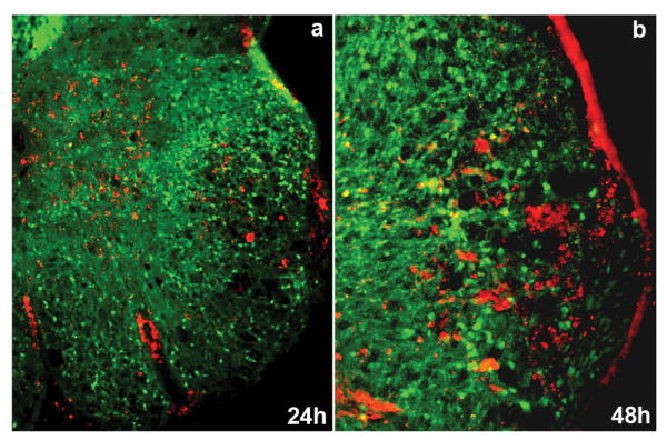
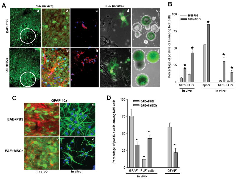
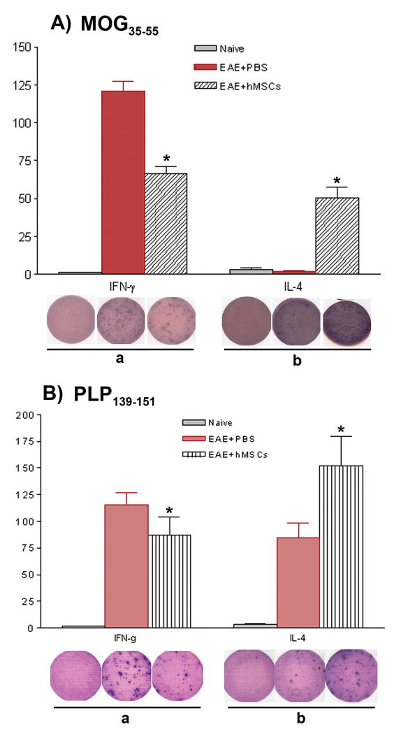
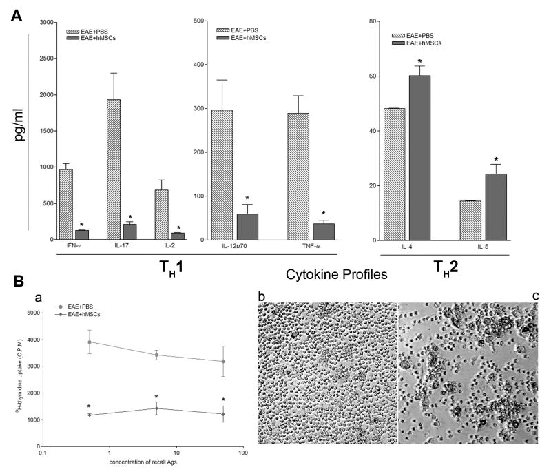
Cell-based therapies are attractive approaches to promote myelin repair. Recent studies demonstrated a reduction in disease burden in mice with experimental allergic encephalomyelitis (EAE) treated with mouse mesenchymal stem cells (MSCs). Here, we demonstrated human bone marrow-derived MSCs (BM-hMSCs) promote functional recovery in both chronic and relapsing-remitting models of mouse EAE, traced their migration into the injured CNS and assayed their ability to modulate disease progression and the host immune response. Injected BM-hMSCs accumulated in the CNS, reduced the extent of damage and increased oligodendrocyte lineage cells in lesion areas. The increase in oligodendrocytes in lesions may reflect BM-hMSC-induced changes in neural fate determination, since neurospheres from treated animals gave rise to more oligodendrocytes and less astrocytes than nontreated neurospheres. Host immune responses were also influenced by BM-hMSCs. Inflammatory T-cells including interferon gamma producing Th1 cells and IL-17 producing Th17 inflammatory cells and their associated cytokines were reduced along with concomitant increases in IL-4 producing Th2 cells and anti-inflammatory cytokines. Together, these data suggest that the BM-hMSCs represent a viable option for therapeutic approaches.


Figures

Figure 1
Figure 1. Intravenous injection of BM-hMSCs ameliorates MOG35-55 and PLP139-151 induced EAE
EAE induced in PLP-EGFP, C57BL/6J and SJL mice by immunization with MOG35-55 and PLP139-151 peptide (a-c). Administration of BM-hMSCs to EGFP transgenic mice at day 16 and day 26 (a), C57BL/6J mice at day 16 (b) or SJL mice at day 16 (c) resulted in improved clinical scores. The clinical scores distributed for each BM-hMSC-treated EGFP transgenic mice at day 45 (d), C57/BL/6J mice at day 45 (e), and in SJL mouse at day 60 (f). Recovery was sustained for up to 70 days the longest time point examined. Arrows indicate days of BM-hMSC injection.
Figure 2
Figure 2. Treatment with BM-hMSCs reduces cellular infiltration and tissue damage in EAE
(A) Low (a,e,c,g) and high (b,f,g,h) power images of H&E and anti-CD45 antibody labeled sections of cervical spinal cord at day 45 post immunization. Carrier treated (d16) EAE mice induced by MOG35-55 contained prominent H&E positive infiltrating cells and large numbers of CD45+ mononuclear leukocytes in lesion areas of white matter (a-d). In animals treated with BM-hMSCs on day 16 when the clinical score was approximately 3.5 and sacrificed on day 45 the numbers of both H&E (f) and anti-CD45 (h) cells were greatly reduced. High magnification images (40X) from marked lesion areas (b,d,f,h). (B) Quantization of the relative proportion of cells in lesion areas in PBS and BM-hMSC treated animals. The number of infiltrating cells is significantly reduced in BM-hMSC treated animals (Mean +/- SD with p <0.05). (C) Myelin staining with Luxol Fast Blue (LFB) at the same time point as A and B. In cervical spinal cord sections from EAE+PBS mice with a score of 3.5 extensive myelin degeneration (d,e) is seen compared to naïve animals (a,b). Animals treated with BM-hMSCs have substantially less myelin loss that than carrier controls (g,h). Similarly, 1μm sections labeled with Toludine Blue (TB) showed increased myelin debris and greater numbers of demyelinated axons in the EAE+PBS mice (f) compared to naïve mice (c). Normalization of cytoarchitecture and reduced myelin debris is evident in lesions from mice treated with BM-hMSCs (i). (D) Comparison of lesion load between BM-MSC treated and non-treated EAE animals and (E) density of myelinated axons in naïve, BM-MSC treated and non-treated animals. Lesion load data was taken from 5 different sections of cervical spinal cord. Axonal counts were taken from lesion areas in the dorsal columns from the same cohort of animals. (N=5) P values determined by two-tailed t test.
Figure 3
Figure 3. BM-hMSCs accumulate in demyelinating lesions in EAE induced by MOG35-55
BM-hMSCs labeled with CMFDA (red) were found in lesion areas of PLP-EGFP transgenic EAE mice at 24h (a) and 48h (b) post-injection.
Figure 4
Figure 4. Treatment with BM-hMSCs increases oligodendrogenesis and decreases astrogliosis in EAE animals
(A) Low numbers of PLP- EGFP+ oligodendrocyte (green) and NG2+ oligodendrocyte precursor cells (red) are present in EAE lesions of carrier treated animals sacrificed at day 45 (a,b). Similarly, cell cultures from lesioned spinal cords (c,d) or neurospheres derived at day 45 from the SVZ of PLP-EGFP EAE mice treated with PBS showed low numbers of oligodendrocyte lineage cells. By contrast, in animals treated with BM-HMSCs on day 16 the numbers of PLP+ cells and NG2+ cells are increased in lesion area, (f,g) in vitro (h,i) and in neurospheres (B) Quantification of relative cell numbers shown in A. The proportion of NG2 cells increases from 5% to 20% in vivo and 2% to 30% in vitro after BM-hMSC treatment. Likewise the proportion of EGFP-PLP+ cells increased from 10% to 57% in vivo and 1% to 14% in vitro after BM-hMSC treatment. The proportion of strongly PLP-EGFP+ neurospheres was significantly increased from 50% to 80% after treatment with BM-hMSCs. (C) Astrogliosis and oligodendrogenesis characterized by increases in GFAP expression (red) and decreases in EGP-PLP expression (green) are prominent in EAE lesions (a) in mice treated with PBS (b) Parallel lesions from animals treated with BM-hMSCs. Note the reduced number of astrocytes (GFAP+) and enhanced oligodendrocytes (PLP-EGFP+ cells). Consistent with reduced astrogliosis in BM-hMSC treated lesions, the proportion of GFAP+ cells in neurosphere cultures from the SVZ of EAE mice treated with BM-hMSCs on day 16 was significantly reduced (d) compared to EAE mice treated with PBS (c). (D) Quantification of the relative cell numbers shown in C. The proportion of GFAP+ cells decreased from 73% to 37% in vivo and 52% to 14% in vitro after BM-hMSC treatment while the number of PLP-EGFP+ cells increased from 10% to 54%. (*) = Significance p<0.05.
Figure 5
Figure 5. BM-hMSC treated EAE mice display reduced frequencies of myelin peptide-specific TH1 but increased frequencies of myelin peptide-specific TH2 cells
106 splenocytes from naïve, PBS-treated or BM-hMSC-treated PLP-EGFP transgenic mice at day 45 post-immunization with MOG35-55-induced EAE (A) or SJL mice at day 60 post-immunization with PLP139-151-induced EAE (B) were stimulated with myelin peptide, then IFN-γ and IL-4 producing T cell ELISOPTS were performed. BM-hMSC-treated mice from both strains displayed decreased frequencies of TH1, but higher frequencies of myelin peptide-specific TH2 compared to PBS-treated controls. The wells shown are representatives from triplicates. Counts represent mean standard deviation from one of three experiments with similar results. (*) = significant difference of p< 0.05.
Figure 6
Figure 6. Differential expression of TH1, TH17 and TH2 cytokine profiles in peripheral lymph organs and suppression of MOG35-55-specific T-cell proliferative responses in BM-hMSC-treated mice with EAE induced by MOG35-55
Cells were isolated from both PBS and BM-hMSC-treated mice at day 16 after immunization and cultured in the presence of MOG35-55 peptide. (A) Responses from the BM-hMSC-treated animals displayed significantly decreased levels of pro-inflammatory cytokines (IFN-γ, IL-17, IL-2, IL-12p70, TNF-α) compared to those from PBS-treated controls. In contrast, the expression of anti-inflammatory cytokines (IL-4, IL-5) was significantly increased in the BM-hMSC-treated group compared to the controls (B). Values are means ± SEM of triplicate cultures. Data represent three separate experiments using spleen cells pooled from three different donors. (*) = significant difference of p< 0.05. (C) Splenic T-cells from control and BM-hMSC-treated EAE mice were cultured with different concentrations of MOG35-55 (0.5 μg/ml, 5 μg/ml, 50 μg/ml) and 3H-thymidine incorporation assessed three days later (a). T cells, at 5ug/ml from BM-hMSC treated mice, showed fewer proliferating colonies (b) than T cells from control mice in response to 5 μg/ml of MOG35-55 (c). Proliferative responses are expressed as mean CPM of at least 3 independent experiments. (*) = significant difference of p< 0.05.

References

    1. Aggarwal S, Pittenger MF. Human mesenchymal stem cells modulate allogeneic immune cell responses. Blood. 2005;105(4):1815–22. - PubMed
    1. Aharonowiz M, Einstein O, Fainstein N, Lassmann H, Reubinoff B, Ben-Hur T. Neuroprotective effect of transplanted human embryonic stem cell-derived neural precursors in an animal model of multiple sclerosis. PLoS ONE. 2008;3(9):e3145. - PMC - PubMed
    1. Alexanian AR. Epigenetic modifiers promote efficient generation of neural-like cells from bone marrow-derived mesenchymal cells grown in neural environment. J Cell Biochem. 2007;100(2):362–71. - PubMed
    1. Ankeny DP, McTigue DM, Jakeman LB. Bone marrow tranplants provide tissue protectionadn directional guidance for axons after contusive spinal cord injuries in rats. Exp Neurology. 2004;190:17–31. - PubMed
    1. Antonysamy MA, Fanslow WC, Fu F, Li W, Qian S, Troutt AB, Thomson AW. Evidence for a role of IL-17 in alloimmunity: a novel IL-17 antagonist promotes heart graft survival. Transplant Proc. 1999;31(1-2):93. - PubMed

Publication types

MeSH terms