. 2009 May 15;75(3):760-73.
doi: 10.1002/prot.22287.

Structural genomics reveals EVE as a new ASCH/PUA-related domain

Affiliations

Structural genomics reveals EVE as a new ASCH/PUA-related domain

Claudia Bertonati et al. Proteins. .

Abstract

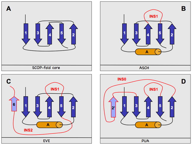
We report on several proteins recently solved by structural genomics consortia, in particular by the Northeast Structural Genomics consortium (NESG). The proteins considered in this study differ substantially in their sequences but they share a similar structural core, characterized by a pseudobarrel five-stranded beta sheet. This core corresponds to the PUA domain-like architecture in the SCOP database. By connecting sequence information with structural knowledge, we characterize a new subgroup of these proteins that we propose to be distinctly different from previously described PUA domain-like domains such as PUA proper or ASCH. We refer to these newly defined domains as EVE. Although EVE may have retained the ability of PUA domains to bind RNA, the available experimental and computational data suggests that both the details of its molecular function and its cellular function differ from those of other PUA domain-like domains. This study of EVE and its relatives illustrates how the combination of structure and genomics creates new insights by connecting a cornucopia of structures that map to the same evolutionary potential. Primary sequence information alone would have not been sufficient to reveal these evolutionary links.


Figures

Fig. 1
Fig. 1. PUA-like domain topologies
In blue and orange we denote strands and helices respectively. Insertions (INS0, INS1, INS2) characteristic of specific domains are marked in red and additional strands (i.e. strands within insertions, namely 6 and 2′) in lavender. The four panels represent: (A) the basic elements (core) of the PUA-like domains topology as reported in SCOP , (B) the ASCH domain architecture , (C) the EVE domain architecture, (D) the architecture of the PUA domains described in the text.
Fig. 2
Fig. 2. Structure-based sequence alignment of 2eve , 2ar1 , 2g2x , 1zce , 2gbs , 2hd9/1wmm - and 2p5d (EVE domains)
The sequence alignments are obtained automatically from pairwise structural alignments between 2eve and the other proteins (Table 2). Each protein is represented by two lines, one giving the sequence, the other the secondary structure assignment (according to Skan ,). Numbers at each end indicate the first and last residue found in the structure, following the PDB sequence numbering for each protein. Below the alignment we highlight the topology of the EVE domains (with strands 1–6, helix A and the two insertions INS1 and INS2). Boxes mark residues that are generally not only conserved in the structural alignment but also in sequence-based alignments from PSI-BLAST and CLUSTALW or Pfam . Conserved residues and numbers reported above the boxes are as in the 2eve PDB file.
Fig. 3
Fig. 3. Ligand binding and surface conservation in the main surface cavity of EVE domains
(A) 2eve structure in cartoon representation (green) with co-crystallized ligands 3[N-morpholino]propane sulfonic acid (MPO) and tris-hydroxymethyl-methyl-ammonium (TRIS) in ball and stick representation (CPK colors). In red (licorice representation) we show conserved residues (Y3, W4, F13, W26, V29, D45, Y50, W142). (B) 2eve molecular surface colored by residue conservation score (as calculated by ConSurf ), conserved residues are in purple, variable residues are in cyan, the orientation of 2eve is as in (A). MPO and TRIS are in ball and stick representation (CPK colors).
Fig. 4
Fig. 4. Structure-based sequence alignment between ASCH and EVE domains
Notations are as in Fig. 2. The alignment is obtained from pairwise structural alignments between 1s04 and the other proteins, except for 2g2x , whose sequence was manually added taking its pairwise structural alignment to 2ar1 as a template (since Skan failed to identify 2g2x as a structural homolog of 1s04). For simplicity, alignments are reported only up to INS1, i.e. the insertion between strand 4 and 5. ASCH sequences are on top and EVE at the bottom. Numbers at each end of a sequence indicate the first and last residues following the PDB sequence numbering for each protein. As in Fig. 3, boxes highlight conserved residues generally indicating not only conservation in the structural alignment but also in sequence-based alignments obtained using PSI-BLAST and CLUSTALW or Pfam . Type and numbers for these residues are as in the PDB sequence of 2eve . Note that in this alignment W26 is slightly misaligned in the EVE domains (compare to Fig. 2) and so is the ASCH motif GxKxxE/T/SxR in the ASCH domains.
Fig. 5
Fig. 5. Virtual complex of 2eve and tRNA from 1j2b
The figure was obtained according to the following procedure: first, 2eve was superimposed on the PUA domain of ArcTGT 1j2b complexed with tRNA (residues 507–582) (using GRASP2 ); then, the 1j2b PUA domain structure was removed while its bound tRNA and 2eve and its ligand (MPO) was kept in place. (A) 2eve structure is cartoon representation (blue), tRNA (CPK colors), MPO (green) are in ball and stick representation. Relevant residues (see text) are in licorice representation (blue). (B) 2eve molecular surface colored by residue conservation score (as calculated by ConSurf ), conserved residues are in purple, variable residues are in cyan, the orientation of 2eve is as in (A). tRNA (CPK colors), MPO (green) are in ball and stick representation.

References

    1. Blundell TL, Mizuguchi K. Structural genomics: an overview. Prog Biophys Mol Biol. 2000;73(5):289–295. - PubMed
    1. Bhattacharya A, Tejero R, Montelione GT. Evaluating protein structures determined by structural genomics consortia. Proteins. 2006 - PubMed
    1. Levitt M. Growth of novel protein structural data. Proc Natl Acad Sci U S A. 2007;104(9):3183–3188. - PMC - PubMed
    1. Berman HM, Battistuz T, Bhat TN, Bluhm WF, Bourne PE, Burkhardt K, Feng Z, Gilliland GL, Iype L, Jain S, Fagan P, Marvin J, Padilla D, Ravichandran V, Schneider B, Thanki N, Weissig H, Westbrook JD, Zardecki C. The Protein Data Bank. Acta Crystallogr D Biol Crystallogr. 2002;58(Pt 61):899–907. - PubMed
    1. Liu J, Montelione GT, Rost B. Novel leverage of structural genomics. Nature Biotechnology. 2007 in press. - PubMed

Publication types

MeSH terms

LinkOut - more resources