. 2009 Feb 24;48(7):1532-42.
doi: 10.1021/bi801942a.

Structure of a SusD homologue, BT1043, involved in mucin O-glycan utilization in a prominent human gut symbiont

Affiliations

Structure of a SusD homologue, BT1043, involved in mucin O-glycan utilization in a prominent human gut symbiont

Nicole Koropatkin et al. Biochemistry. .

Abstract

Mammalian distal gut bacteria have an expanded capacity to utilize glycans. In the absence of dietary sources, some species rely on host-derived mucosal glycans. The ability of Bacteroides thetaiotaomicron, a prominent human gut symbiont, to forage host glycans contributes to both its ability to persist within an individual host and its ability to be transmitted naturally to new hosts at birth. The molecular basis of host glycan recognition by this species is still unknown but likely occurs through an expanded suite of outermembrane glycan-binding proteins that are the primary interface between B. thetaiotaomicron and its environment. Presented here is the atomic structure of the B. thetaiotaomicron protein BT1043, an outer membrane lipoprotein involved in host glycan metabolism. Despite a lack of detectable amino acid sequence similarity, BT1043 is a structural homologue of the B. thetaiotaomicron starch-binding protein SusD. Both structures are dominated by tetratrico peptide repeats that may facilitate association with outer membrane beta-barrel transporters required for glycan uptake. The structure of BT1043 complexed with N-acetyllactosamine reveals that recognition is mediated via hydrogen bonding interactions with the reducing end of beta-N-acetylglucosamine, suggesting a role in binding glycans liberated from the mucin polypeptide. This is in contrast to CBM 32 family members that target the terminal nonreducing galactose residue of mucin glycans. The highly articulated glycan-binding pocket of BT1043 suggests that binding of ligands to BT1043 relies more upon interactions with the composite sugar residues than upon overall ligand conformation as previously observed for SusD. The diversity in amino acid sequence level likely reflects early divergence from a common ancestor, while the unique and conserved alpha-helical fold the SusD family suggests a similar function in glycan uptake.


Figures
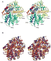
Figure 1
Figure 1. The 2.0 Å structure of selenomethionine-substituted BT1043
(A) Cartoon representation of the apo BT1043, colored blue to red from N- to C-terminus. The N-acetyl lactosamine ligand (magenta spheres) has been modeled in to show the location of the glycan-binding pocket. The 18 α-helices and 6 β-strands are labeled as shown; 310 helices are not labeled. Four helix-turn-helix pairs, or tetratrico peptide repeats (TPRs), are labeled with α1 & α4 as TPR1, α5 & α6 as TPR2, α7 & α8 as TPR3, and α12 & α13 as TPR4. (B) Overlay of BT1043, shown in blue, SusD (PDB 3CK7), shown in green, and BT3984 (PDB 3CGH) shown in red, in a similar view as in panel A to highlight to similarities in the TPR domain.
Figure 2
Figure 2. N-acetyl lactosamine (LacNAc) binding in the 2.8Å structure of BT1043
(A) Omit map (3σ) of native BT1043 complexed with LacNAc. The reducing O1 oxygen of N-acetylglucosamine (GlcNAc) and the non-reducing O4 oxygen of galactose are labeled, along with the α-helices that shape the glycan-binding site. (B.) Close-up view of LacNAc binding to BT1043. Coordinating and nearby residues are displayed, with distances labeled in angstroms (Å).
Figure 3
Figure 3. Overlay of the SusD and BT1043 glycan-binding pockets
(A). Close-up view of the amino acids lining the glycan binding pocket of BT1043 (blue) with that of SusD (pink). Starch-binding residues in SusD are overlayed with the equivalent residues in BT1043. The ribbon representation of the glycan binding pocket is shown to highlight the similarities in the overall shape and size of site, despite differences in amino acid sequence. (B) Close-up view of the BT1043 (blue-purple) LacNAc binding site with the equivalent glycan-binding site in BT3984 (yellow-orange).
Figure 4
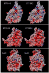
Figure 4. Comparison of glycan binding pockets of BT1043, BT3984, and SusD
Shown here are stereo pairs of surface representations generated in the program CHIMERA (46) of the glycan binding surfaces of these three polysaccaride binding proteins colored according to the electrostatic potential (red for negative and blue for positive) calculated in the program GRASP (47) using the DelPhi (48) algorithm. The ball and stick figure in the BT1043 model represents the structure of the bound LacNAc and α-cyclodextrin in the case of the SusD surface representation.
None

References

    1. Savage DC. Microbial ecology of the gastrointestinal tract. Annu Rev Microbiol. 1977;31:107–133. - PubMed
    1. Ley RE, Hamady M, Lozupone C, Turnbaugh PJ, Ramey RR, Bircher JS, Schlegel ML, Tucker TA, Schrenzel MD, Knight R, Gordon JI. Evolution of mammals and their gut microbes. Science. 2008;320:1647–1651. - PMC - PubMed
    1. Palmer C, Bik EM, Digiulio DB, Relman DA, Brown PO. Development of the Human Infant Intestinal Microbiota. PLoS Biol. 2007;5:e177. - PMC - PubMed
    1. Shroff KE, Meslin K, Cebra JJ. Commensal enteric bacteria engender a self-limiting humoral mucosal immune response while permanently colonizing the gut. Infect Immun. 1995;63:3904–3913. - PMC - PubMed
    1. Mazmanian SK. Capsular polysaccharides of symbiotic bacteria modulate immune responses during experimental colitis. J Pediatr Gastroenterol Nutr. 2008;46(Suppl 1):E11–12. - PubMed

Publication types

LinkOut - more resources