. 2009 Jul;15(7):1797-805.
doi: 10.1089/ten.tea.2008.0364.

Neural stem cell- and Schwann cell-loaded biodegradable polymer scaffolds support axonal regeneration in the transected spinal cord

Affiliations

Neural stem cell- and Schwann cell-loaded biodegradable polymer scaffolds support axonal regeneration in the transected spinal cord

Heather E Olson et al. Tissue Eng Part A. 2009 Jul.

Abstract

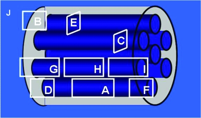
Biodegradable polymer scaffolds provide an excellent approach to quantifying critical factors necessary for restoration of function after a transection spinal cord injury. Neural stem cells (NSCs) and Schwann cells (SCs) support axonal regeneration. This study examines the compatibility of NSCs and SCs with the poly-lactic-co-glycolic acid polymer scaffold and quantitatively assesses their potential to promote regeneration after a spinal cord transection injury in rats. NSCs were cultured as neurospheres and characterized by immunostaining for nestin (NSCs), glial fibrillary acidic protein (GFAP) (astrocytes), betaIII-tubulin (immature neurons), oligodendrocyte-4 (immature oligodendrocytes), and myelin oligodendrocyte (mature oligodendrocytes), while SCs were characterized by immunostaining for S-100. Rats with transection injuries received scaffold implants containing NSCs (n=17), SCs (n=17), and no cells (control) (n=8). The degree of axonal regeneration was determined by counting neurofilament-stained axons through the scaffold channels 1 month after transplantation. Serial sectioning through the scaffold channels in NSC- and SC-treated groups revealed the presence of nestin, neurofilament, S-100, and betaIII tubulin-positive cells. GFAP-positive cells were only seen at the spinal cord-scaffold border. There were significantly more axons in the NSC- and SC- treated groups compared to the control group. In conclusion, biodegradable scaffolds with aligned columns seeded with NSCs or SCs facilitate regeneration across the transected spinal cord. Further, these multichannel biodegradable polymer scaffolds effectively serve as platforms for quantitative analysis of axonal regeneration.


Figures

FIG. 1.
FIG. 1.
Characterization of NSCs and SCs in vitro. Phase contrast image of a neurosphere in culture (A). Confocal microscopy images of neurospheres stained with Nestin (B), βIII-tubulin (C), GFAP (D), O4 (E), and MOSP (F). Bisbenzimide (blue) was used to counterstain the nuclei of all cells in (B–F). Phase contrast image of SCs in culture (G). Fluorescent microscopy image of SCs stained with S100 (H). Magnification: 100 × (A–F); 200 × (G, H). Color images available online at www.liebertonline.com/ten.
FIG. 2.
FIG. 2.
NSC and SC survival/morphology when loaded into PLGA scaffolds in vitro. The 85:15 PLGA scaffolds were 2 mm in length, 3 mm in diameter, and contained 660-μm-diameter longitudinal channels (A). Autofluorescence of the scaffolds occurred. Neurospheres or SCs, stained with bisbenzimide 33342 (blue), were evenly distributed in the channels after 2 h in culture (B, D), and proliferated to fill the channels after 65 h in culture (C, E), respectively. NSCs retained their ability to differentiate when cultured in the scaffold (F) (taken 24 h postseeding). All images were taken transversely at the end of the scaffold channels using a confocal microscope. Magnification: 25× (A); 50 × (B–E); 100 × (F). Color images available online at www.liebertonline.com/ten.
FIG. 3.
FIG. 3.
Characterization of cells within the scaffolds 1 month after transplantation into the transected cord. Confocal (A–C, E, G–I) and fluorescent (D, F) microscopy images of nestin (A), neurofilament (NF) (B, E, G–I), GFAP (B, D, F, G–I), βIII tubulin (C), Gal C (C), APC (D, F), stained cells in NSC-loaded (A–D), and SC-loaded (E–I) channels. Transverse sections: (C, E). Longitudinal sections: (A, B, D, F–I). (J) Schematic representation of the location at which images (A–I) were taken in the scaffold. In image (D) asterisk (*) denotes the border between the spinal cord and scaffold. Magnification: 200 × (F–I); 400 × (A, B, D, E); 630 × (C). Color images available online at www.liebertonline.com/ten.
FIG. 3.
FIG. 3.
Characterization of cells within the scaffolds 1 month after transplantation into the transected cord. Confocal (A–C, E, G–I) and fluorescent (D, F) microscopy images of nestin (A), neurofilament (NF) (B, E, G–I), GFAP (B, D, F, G–I), βIII tubulin (C), Gal C (C), APC (D, F), stained cells in NSC-loaded (A–D), and SC-loaded (E–I) channels. Transverse sections: (C, E). Longitudinal sections: (A, B, D, F–I). (J) Schematic representation of the location at which images (A–I) were taken in the scaffold. In image (D) asterisk (*) denotes the border between the spinal cord and scaffold. Magnification: 200 × (F–I); 400 × (A, B, D, E); 630 × (C). Color images available online at www.liebertonline.com/ten.
FIG. 4.
FIG. 4.
Neurofilament staining of axons in transverse sections through the scaffold after 1 month in vivo. Fluorescent microscopy image of one transverse section of a scaffold (loaded with NSCs) stained with an antibody against neurofilament with the region in which axons were counted encircled by a white line (A). Transverse sections of one channel from the NSC group (B), one from the SC group (C), and one from the control group (D) stained for neurofilament using an AEC chromogen. Magnification: 25× (A); 100× (B–D). Dot plots of axonal counts per cord in the NSC-, SC-, and control-treated groups (E). Data points represent individual animals. 95% Confidence interval was used for each group. *Medians vary significantly (p < 0.05) (p = 0.0162 using the Kruskal–Wallis test) when compared to the control group. Color images available online at www.liebertonline.com/ten.

References

    1. Quencer R.M. Bunge R.P. The injured spinal cord: imaging, histopathologic clinical correlates, and basic science approaches to enhancing neural function after spinal cord injury. Spine. 1996;21:2064. - PubMed
    1. Bodley R. Imaging in chronic spinal cord injury—indications and benefits. Eur J Radiol. 2002;42:135. - PubMed
    1. Friedman J.A. Windebank A.J. Moore M.J. Spinner R.J. Currier B.L. Yaszemski M.J. Biodegradable polymer grafts for surgical repair of the injured spinal cord. Neurosurgery. 2002;51:742. - PubMed
    1. Novikova L.N. Novikov L.N. Kellerth J.O. Biopolymers and biodegradable smart implants for tissue regeneration after spinal cord injury. Curr Opin Neurol. 2003;16:711. - PubMed
    1. Moore M.J. Friedman J.A. Lewellyn E.B. Mantila S.M. Krych A.J. Ameenuddin S. Knight A.M. Lu L. Currier B.L. Spinner R.J. Marsh R.W. Windebank A.J. Yaszemski M.J. Multiple-channel scaffolds to promote spinal cord axon regeneration. Biomaterials. 2006;27:419. - PubMed

Publication types

MeSH terms