. 2009 Jun;22(3):307-18.
doi: 10.1111/j.1755-148X.2009.00551.x. Epub 2009 Feb 3.

Mitf dosage as a primary determinant of melanocyte survival after ultraviolet irradiation

Affiliations

Mitf dosage as a primary determinant of melanocyte survival after ultraviolet irradiation

Thomas J Hornyak et al. Pigment Cell Melanoma Res. 2009 Jun.

Abstract

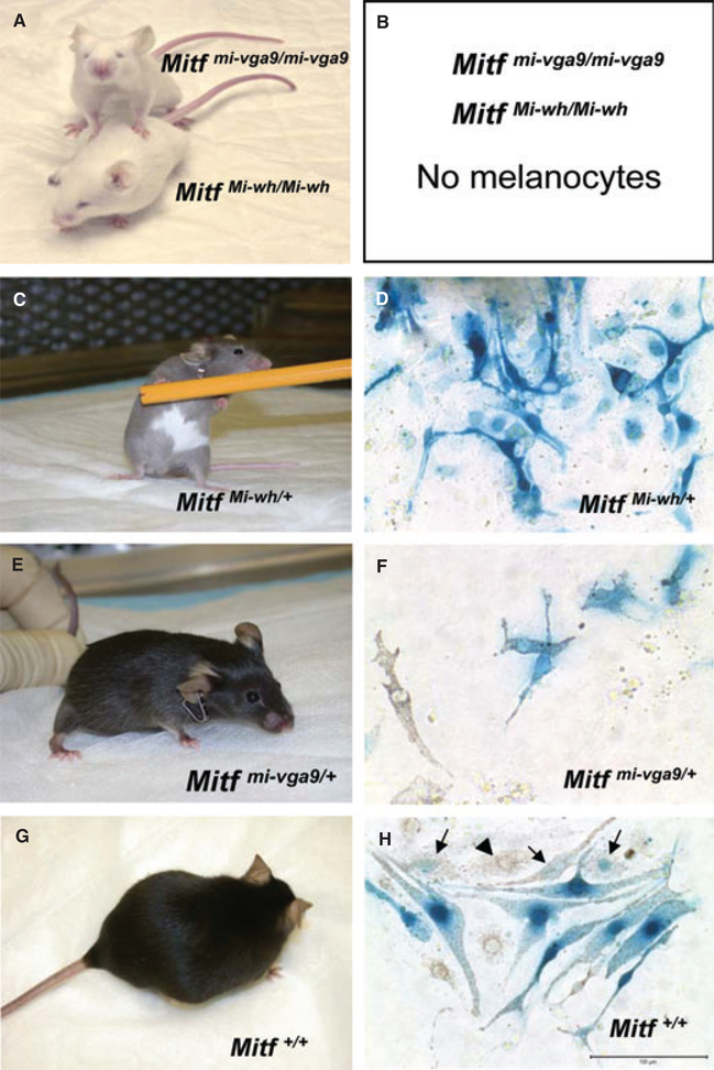
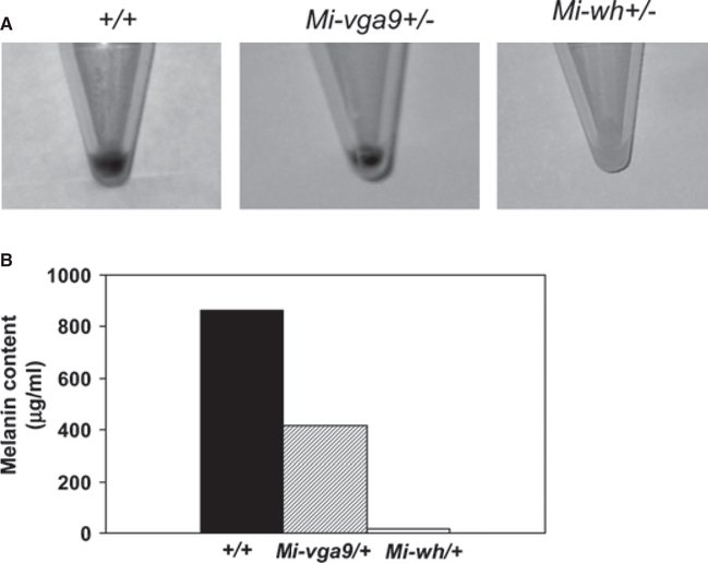
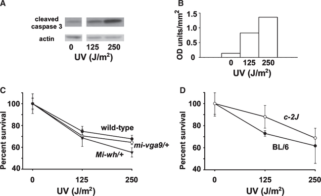
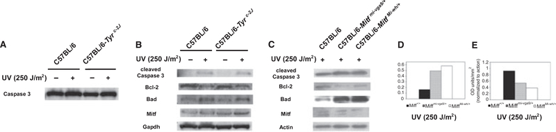
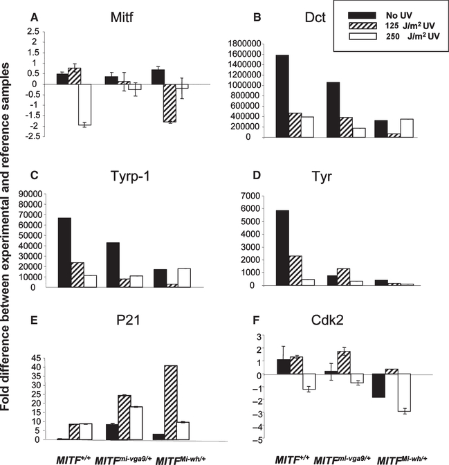
Microphthalmia-associated transcription factor (Mitf) is essential for melanocyte development and function and regulates anti-apoptotic Bcl2 expression. We hypothesized that cellular deficiency of Mitf can influence melanocyte survival in response to ultraviolet (UV) radiation. Primary melanocyte cultures were prepared from neonatal wild-type mice and congenic animals heterozygous for Mitf mutations Mitf (mi-vga9/+) and Mitf(Mi-wh/+) and exposed to UV irradiation. Wild-type melanocytes were more resistant to UV-induced apoptosis than melanocytes partially deficient in Mitf activity, as determined by relative levels of intracellular melanin and relative activation of Mitf target genes Tyr, Tyrp1, Dct, and Cdk2. Comparative experiments with wild-type cells and congenic albino melanocytes demonstrated that these differences are not due to differences in melanin content, implicating Mitf as a primary determinant of UV-dependent melanocyte survival. Mitf activity correlated directly with resistance to UV-induced apoptosis in melanocytes. Mitf was important not only for regulating the expression of anti-apoptotic Bcl-2 following UV irradiation, but also the expression of the pro-apoptotic BH3-only Bad protein and activation of the extrinsic apoptotic pathway. Hence, Mitf is a multifaceted regulator of UV-induced apoptosis in melanocytes.


Figures

Figure 1.
Figure 1.
Phenotype of Mitf mutant mice and their melanocytes. (A) C57BL/6J-MitfMi-wh/Mi-wh and C57BL/6J-Mitfmi-vga9/mi-vga9 homozygous mutant mice. The homozygous mutants lack melanocytes, have microphthalmia (small eyes) and are deaf due to defective melanocyte development. (B) No melanocytes can be isolated from (A). (C) C57BL/6J-MitfMi-wh/+ heterozygous mouse. The Mi-wh mutation in Mitf has a dominant negative effect in vitro and a semi-dominant phenotype in vivo, resulting in grey coat color and large belly spot. (D) Cultured C57BL/6J-MitfMi-wh/+ melanocytes expressing [Tg]Dct-lacZ transgene and stained with X-gal. (E) C57BL/6J Mitfmi-vga9/+ heterozygous mouse. (F) Cultured C57BL/6J-Mitfvga–9/+ melanocytes expressing [Tg]Dct-lacZ transgene and stained with X-gal. (G) C57BL/6 mouse with wild type Mitf. (H) Cultured C57BL/6J wild-type melanocytes expressing [Tg]Dct-lacZ transgene and stained with X-gal. Arrows indicate melanin-containing cells that are losing their [Tg]Dct-lacZ expression. Arrowhead indicates a melanin-containing melanocyte no longer expressing [Tg]Dct-lacZ. All melanocyte cultures were stained for β-galactosidase expression with X-gal (blue). Bar = 100 μm.
Figure 2.
Figure 2.
Melanin content of primary melanocyte cultures. Equal numbers (1 × 10⁶) of cells from wild-type C57BL/6J, C57BL/6J-Mitfmi-vga9/+, and C57BL/6J-MitfMi-wh/+ mice were pelleted (A) and melanin extracted. The melanin content of the cells is shown in (B).
Figure 3.
Figure 3.
Dose response of melanocytes to UV radiation. Primary cultures of melanocytes and melanoblasts from neonatal mice were exposed in serum-free PBS to 0, 125 or 250 J/m2 UV radiation. After washing, fresh media was added and the culture continued for 24 h at 37°C after which the cells were lysed for determination of protein expression with Western blotting (A, B) or utilized in the MTS assay for determination of relative viable cell number. (A) Expression of cleaved caspase 3 in primary C57BL/6 wild-type melanocytes following exposure to increasing doses of UV. β-actin was used as a loading control. (B) Densitometry analysis of (A), with cleaved caspase 3 normalized to actin. (C) Viability of melanocyte cultures from C57BL/6 and congenic C57BL/6J-MitfMi-wh/+ and C57BL/6J-Mitfmi-vga9/+ neonatal mice following irradiation with increasing doses of UV, measured with the MTS assay. Viability decreases as a function of increased Mitf mutational severity (wild-type >Mi-wh/+ > mi-vga9/+). (D) Viability of melanocyte cultures from C57BL/6 and congenic albino C57BL/6J-Tyrc−2J neonatal mice following irradiation with increasing doses of UV, measured with the MTS assay. No significant difference was observed between the viability of albino and pigmented melanocytes following UV irradiation. Error bars represent standard error of the mean.
Figure 4.
Figure 4.
Effect of UV on apoptosis in primary wild-type, albino, and Mitf-deficient melanocytes. Primary cultures of C57BL/6, C57BL/6-Tyrc−2J, C57BL/6-Mitfmi-vga9/+, and C57BL/6-MitfMi-wh/+ melanocytes were exposed to 0 or 250 J/m2 UV radiation. Expression of apoptotic and anti-apoptotic proteins was studied using Western blotting from cell lysates collected 24 h later. (A) Expression of caspase 3 by C57BL/6 and C57BL/6-Tyrc−2J melanocytes. (B) Expression of apoptosis-related proteins caspase 3 (cleaved), Bcl-2, and Bad in irradiated and unirradiated pigmented C57BL/6 and albino C57BL/6-Tyrc−2J primary melanocytes. (C) Expression of apoptosis-related proteins in irradiated C57BL/6, C57BL/6-Mitfmi-vga9/+, and C57BL/6-MitfMi-wh/+ primary melanocytes. (D) Quantification of cleaved caspase 3 expression in irradiated C57BL/6, C57BL/6-Mitfmi-vga9/+, and C57BL/6-MitfMi-wh/+ primary melanocytes in (C). (E) Ratios of quantified Bcl-2 and Bad expression (Bcl-2:Bad ratio) from irradiated C57BL/6, C57BL/6-Mitfmi-vga9/+, and C57BL/6-MitfMi-wh/+ primary melanocytes in (C).
Figure 5.
Figure 5.
Real-time PCR analysis of RNA from melanocyte cultures. Cultures of melanocytes from the genotypes shown in Figure 1 were subjected to 0, 125 J/m2 or 500 J/m2 UV radiation and their RNA harvested 24 h later as described in the Methods. Data are expressed as the fold difference in gene expression between the experimental sample and a reference sample standard, as described in the Methods, using β-actin as an internal control.

Similar articles

Cited by

References

    1. Abdel-Malek ZA, Scott MC, Furumura M, Lamoreux ML, Ollmann M, Barsh GS, and Hearing VJ (2001). The melano-cortin 1 receptor is the principal mediator of the effects of agouti signaling protein on mammalian melanocytes. J. Cell Sci 114, 1019–1024. - PubMed
    1. Aragane Y, Kulms D, Metze D, Wilkes G, Poppelmann B, Luger TA, and Schwarz T (1998). Ultraviolet light induces apoptosis via direct activation of CD95 (Fas/APO-1) independently of its ligand CD95L. J. Cell Biol 140, 171–182. - PMC - PubMed
    1. Bentley NJ, Eisen T, and Goding CR (1994). Melanocyte-specific expression of the human tyrosinase promoter: activation by the microphthalmia gene product and role of the initiator. Mol. Cell. Biol 14, 7996–8006. - PMC - PubMed
    1. Bismuth K, Skuntz S, Hallsson JH, Pak E, Dutra AS, Steingrimsson E, and Arnheiter H (2008). An unstable targeted allele of the mouse Mitf gene with a high somatic and germline reversion rate. Genetics 178, 259–272. - PMC - PubMed
    1. Bivik CA, Andersson EB, and Rosdahl IK (2005). Wavelength-specific effects on UVB-induced apoptosis in melanocytes. A study of Bcl-2/Bax expression and keratinocyte rescue effects. Melanoma Res. 15, 7–13. - PubMed

Publication types

Substances