Review
. 2009 May;10(5):461-71.
doi: 10.1111/j.1600-0854.2009.00885.x. Epub 2009 Jan 24.

Basal body assembly in ciliates: the power of numbers

Affiliations
Review

Basal body assembly in ciliates: the power of numbers

Chad G Pearson et al. Traffic. 2009 May.

Abstract

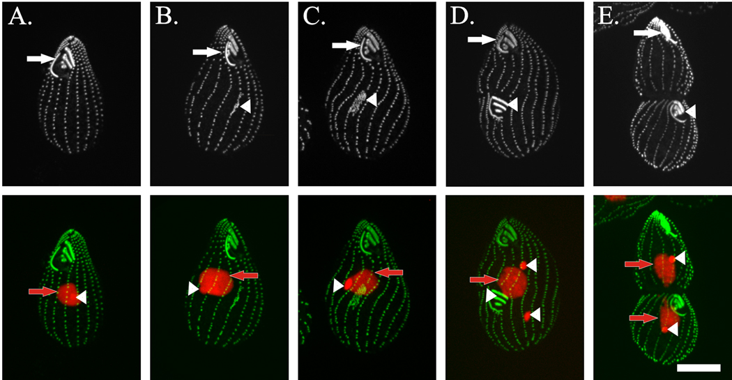
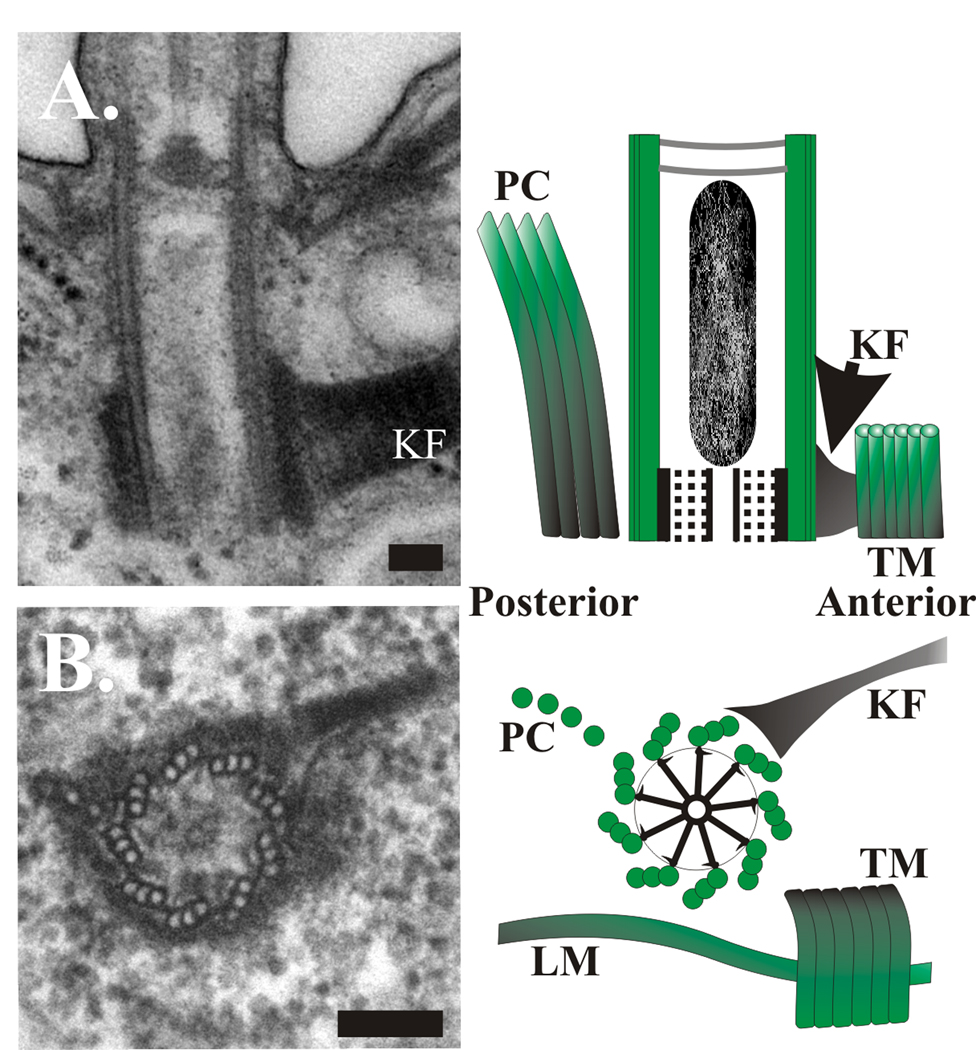
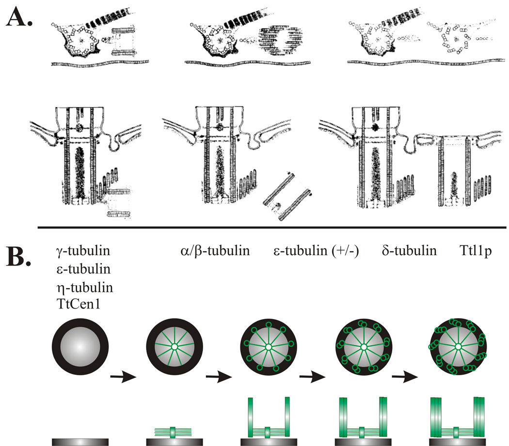
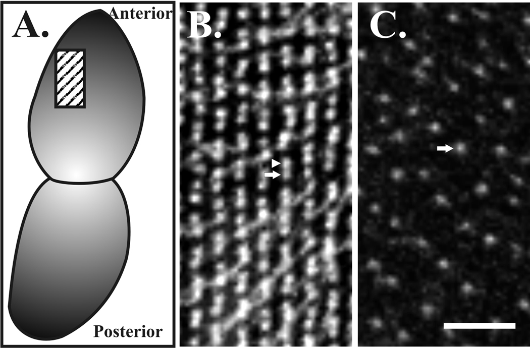
Centrioles perform the dual functions of organizing both centrosomes and cilia. The biogenesis of nascent centrioles is an essential cellular event that is tightly coupled to the cell cycle so that each cell contains only two or four centrioles at any given point in the cell cycle. The assembly of centrioles and their analogs, basal bodies, is well characterized at the ultrastructural level whereby structural modules are built into a functional organelle. Genetic studies in model organisms combined with proteomic, bioinformatic and identifying ciliary disease gene orthologs have revealed a wealth of molecules requiring further analysis to determine their roles in centriole duplication, assembly and function. Nonetheless, at this stage, our understanding of how molecular components interact to build new centrioles and basal bodies is limited. The ciliates, Tetrahymena and Paramecium, historically have been the subject of cytological and genetic study of basal bodies. Recent advances in the ciliate genetic and molecular toolkit have placed these model organisms in a favorable position to study the molecular mechanisms of centriole and basal body assembly.


Figures

Figure 1
Figure 1. New basal body formation during the late stages of Tetrahymena cell division
Tetrahymena thermophila cells with basal bodies labeled by staining with anti-centrin (upper panels) and combined basal bodies (anti-centrin (green)) and DNA (Hoerscht 33342 (red)) (lower panels). (A) Interphase cell with relatively uniform basal body spacing along the ciliary rows. Arrow denotes mature oral apparatus at the cell anterior. DNA staining shows both the larger macronucleus (red arrow) and the smaller, dense micronucleus (white arrowhead). (B) Basal bodies in the ciliary rows increase in density as a result of new basal body assembly. The development of the oral primordium (stomatogenesis) is initiated (arrowhead) at the cell median in preparation for cell division. (C) New basal body assembly occurs at ciliary rows (most of the new basal body assembly is positioned immediately posterior to the division plane) and the oral primordium. The micronucleus begins to elongate into a prolate spheroid or rugby ball shape. (D) The oral primordium organizes into the three membranelles and the crescent shaped undulating membrane. The micronucleus has divided. (E) The division plan is evident with both the micronucleus and macronucleus separated between the mother and daughter cells. Scale bar, 10 µm.
Figure 2
Figure 2. Tetrahymena basal body structure
(A) Longitudinal thin section EM through a single basal body showing basal body cylinder structure and kinetodesmal fiber (KF). The cartoon identifies a longitudinal basal body section with accessory structures. The cartwheel structure is at the base of the organelle. Several of the accessory structures surrounding the basal bodies are indicated. (B) Cross thin section EM through the basal body structure, kinetodesmal fiber, and post-ciliary microtubles. The cartoon reveals a cross section through the cartwheel structure and several of the accessory structures surrounding basal bodies. Labeled structures are as follows: LM, longitudinal microtubule. PC, post-ciliary microtubule bands. KF, kinetodesmal fibers. TM, transverse microtubules. The anterior and posterior positioning relative to the cellular geometry is indicated. Scale bars, 100 nm.
Figure 3
Figure 3. Stages of early basal body assembly in Tetrahymena
Drawing depicts longitudinal sections of basal body formation. Basal bodies form on the anterior side (respective to the cellular geometry) of the parent organelle. Nascent assembly is initiated at the proximal end and perpendicular to the existing parent basal body to form a short pro-basal body. The pro-basal body then separates and tilts parallel to the parent organelle and becomes inserted into the plasma membrane (figure courtesy of The Journal of Cell Biology). Stages of early basal body assembly. (A) Drawing depicts longitudinal sections of basal body formation in Tetrahymena. Basal bodies form on the anterior side (respective to the cellular geometry) of the parent organelle. Nascent assembly is initiated at the proximal end and perpendicular to the existing parent basal body to form a short pro-basal body. The pro-basal body then separates and tilts parallel to the parent organelle and becomes inserted into the plasma membrane (permission from The Journal of Cell Biology). (B) Cartoon depicts the steps to new basal body assembly based on combined studies from Tetrahymena and Paramecium. Assembly begins with the formation of the generative disk, followed by cartwheel assembly, the first structural evidence for nine-fold symmetry. Singlet microtubules are assembled at the ends of each cartwheel spoke followed by the B- and C-tubules to generate doublet and triplet microtubules, respectively. Genes involved in each transition stage are indicated, based upon the molecular components described in this review. (+/−) indicates partial ɛ-tubulin depletion. Note that this is not an exhaustive list of the known molecular components. Top panels, cross section. Bottom panels, longitudinal section.
Figure 4
Figure 4. Loss of basal body duplication in Paramecium cells
(A) The cartoon represents a dividing Paramecium cell at the stages visualized and box indicates the anterior region of the dorsal side taken from both wild-type (B) and ɛ-tubulin depleted (C) cells. Basal bodies (and some cilia) are stained with anti-α-tubulin antibodies (ID5). (B) In wild-type cells, basal body doublets are easily visualized in close proximity to each other. Each doublet contains a mature basal body (arrow, posterior with associated cilia detectable at some basal bodies) and an immature basal body (arrowhead, anterior). (C) Cells depleted of ɛ-tubulin by RNAi for 72 hours do not have pairs of duplicated basal bodies (arrow). Images were kindly provided by Dr. Pascale Dupuis-Williams (Université Evry Val d’Essonne, France). Scale bar, 2 µm.

Similar articles

Cited by

References

    1. Boveri T. Germany: Jena; 1914. Zur frage der entstehung maligner tumoren.
    1. Wilson E. 3rd ed. New York: Macmillan; 1925. The Cell in Development and Heredity.
    1. Bornens M, Azimzadeh J. Origin and evolution of the centrosome. Advances in experimental medicine and biology. 2007;607:119–129. - PubMed
    1. Basto R, Lau J, Vinogradova T, Gardiol A, Woods CG, Khodjakov A, Raff JW. Flies without centrioles. Cell. 2006;125(7):1375–1386. - PubMed
    1. Marshall WF, Nonaka S. Cilia: tuning in to the cell's antenna. Curr Biol. 2006;16(15):R604–R614. - PubMed

Publication types

LinkOut - more resources