Pseudomonas aeruginosa vesicles associate with and are internalized by human lung epithelial cells
- PMID: 19192306
- PMCID: PMC2653510
- DOI: 10.1186/1471-2180-9-26
Pseudomonas aeruginosa vesicles associate with and are internalized by human lung epithelial cells
Abstract
Background: Pseudomonas aeruginosa is the major pathogen associated with chronic and ultimately fatal lung infections in patients with cystic fibrosis (CF). To investigate how P. aeruginosa-derived vesicles may contribute to lung disease, we explored their ability to associate with human lung cells.
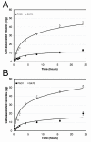
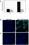
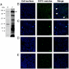
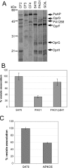
Results: Purified vesicles associated with lung cells and were internalized in a time- and dose-dependent manner. Vesicles from a CF isolate exhibited a 3- to 4-fold greater association with lung cells than vesicles from the lab strain PAO1. Vesicle internalization was temperature-dependent and was inhibited by hypertonic sucrose and cyclodextrins. Surface-bound vesicles rarely colocalized with clathrin. Internalized vesicles colocalized with the endoplasmic reticulum (ER) marker, TRAPalpha, as well as with ER-localized pools of cholera toxin and transferrin. CF isolates of P. aeruginosa abundantly secrete PaAP (PA2939), an aminopeptidase that associates with the surface of vesicles. Vesicles from a PaAP knockout strain exhibited a 40% decrease in cell association. Likewise, vesicles from PAO1 overexpressing PaAP displayed a significant increase in cell association.
Conclusion: These data reveal that PaAP promotes the association of vesicles with lung cells. Taken together, these results suggest that P. aeruginosa vesicles can interact with and be internalized by lung epithelial cells and contribute to the inflammatory response during infection.
Figures


Similar articles
-
Pseudomonas aeruginosa Leucine Aminopeptidase Influences Early Biofilm Composition and Structure via Vesicle-Associated Antibiofilm Activity.mBio. 2019 Nov 19;10(6):e02548-19. doi: 10.1128/mBio.02548-19. mBio. 2019. PMID: 31744920 Free PMC article.
-
Purification of outer membrane vesicles from Pseudomonas aeruginosa and their activation of an IL-8 response.Microbes Infect. 2006 Aug;8(9-10):2400-8. doi: 10.1016/j.micinf.2006.05.001. Epub 2006 Jun 5. Microbes Infect. 2006. PMID: 16807039 Free PMC article.
-
Occurrence of hypermutable Pseudomonas aeruginosa in cystic fibrosis patients is associated with the oxidative stress caused by chronic lung inflammation.Antimicrob Agents Chemother. 2005 Jun;49(6):2276-82. doi: 10.1128/AAC.49.6.2276-2282.2005. Antimicrob Agents Chemother. 2005. PMID: 15917521 Free PMC article.
-
Pseudomonas aeruginosa chromosomal beta-lactamase in patients with cystic fibrosis and chronic lung infection. Mechanism of antibiotic resistance and target of the humoral immune response.APMIS Suppl. 2003;(116):1-47. APMIS Suppl. 2003. PMID: 14692154 Review.
-
Microevolution of Pseudomonas aeruginosa to a chronic pathogen of the cystic fibrosis lung.Curr Top Microbiol Immunol. 2013;358:91-118. doi: 10.1007/82_2011_199. Curr Top Microbiol Immunol. 2013. PMID: 22311171 Review.
Cited by
-
Mechanisms of outer membrane vesicle entry into host cells.Cell Microbiol. 2016 Nov;18(11):1508-1517. doi: 10.1111/cmi.12655. Epub 2016 Sep 16. Cell Microbiol. 2016. PMID: 27529760 Free PMC article. Review.
-
Stress-induced outer membrane vesicle production by Pseudomonas aeruginosa.J Bacteriol. 2013 Jul;195(13):2971-81. doi: 10.1128/JB.02267-12. Epub 2013 Apr 26. J Bacteriol. 2013. PMID: 23625841 Free PMC article.
-
Abiotic stressors impact outer membrane vesicle composition in a beneficial rhizobacterium: Raman spectroscopy characterization.Sci Rep. 2020 Dec 4;10(1):21289. doi: 10.1038/s41598-020-78357-4. Sci Rep. 2020. PMID: 33277560 Free PMC article.
-
Role of Legionella pneumophila outer membrane vesicles in host-pathogen interaction.Front Microbiol. 2023 Sep 25;14:1270123. doi: 10.3389/fmicb.2023.1270123. eCollection 2023. Front Microbiol. 2023. PMID: 37817751 Free PMC article. Review.
-
Extracellular Vesicles as Biomarkers in Infectious Diseases.Biology (Basel). 2025 Feb 11;14(2):182. doi: 10.3390/biology14020182. Biology (Basel). 2025. PMID: 40001950 Free PMC article. Review.
References
-
- Yoon SS, Hennigan RF, Hilliard GM, Ochsner UA, Parvatiyar K, Kamani MC, Allen HL, DeKievit TR, Gardner PR, Schwab U, Rowe JJ, Iglewski BH, McDermott TR, Mason RP, Wozniak DJ, Hancock RE, Parsek MR, Noah TL, Boucher RC, Hassett DJ. Pseudomonas aeruginosa anaerobic respiration in biofilms: relationships to cystic fibrosis pathogenesis. Developmental cell. 2002;3(4):593–603. doi: 10.1016/S1534-5807(02)00295-2. - DOI - PubMed
-
- Worlitzsch D, Tarran R, Ulrich M, Schwab U, Cekici A, Meyer KC, Birrer P, Bellon G, Berger J, Weiss T, Botzenhart K, Yankaskas JR, Randell S, Boucher RC, Doring G. Effects of reduced mucus oxygen concentration in airway Pseudomonas infections of cystic fibrosis patients. The Journal of clinical investigation. 2002;109(3):317–325. - PMC - PubMed
-
- Malloy JL, Veldhuizen RA, Thibodeaux BA, O'Callaghan RJ, Wright JR. Pseudomonas aeruginosa protease IV degrades surfactant proteins and inhibits surfactant host defense and biophysical functions. American journal of physiology. 2005;288(2):L409–418. - PubMed
Publication types
MeSH terms
Substances
Grants and funding
LinkOut - more resources
Full Text Sources
Other Literature Sources
Medical
Miscellaneous

