. 2009 Feb 3:6:10.
doi: 10.1186/1742-4690-6-10.

Rev and Rex proteins of human complex retroviruses function with the MMTV Rem-responsive element

Affiliations

Rev and Rex proteins of human complex retroviruses function with the MMTV Rem-responsive element

Jennifer A Mertz et al. Retrovirology. .

Abstract

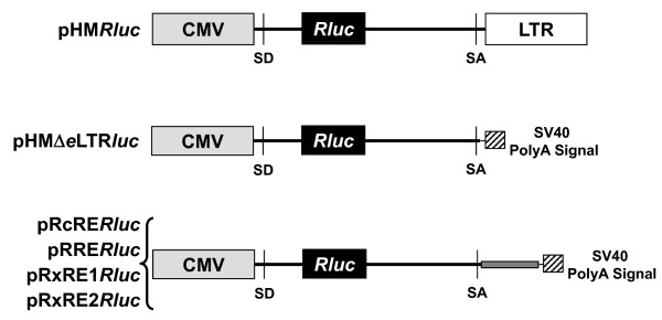
Background: Mouse mammary tumor virus (MMTV) encodes the Rem protein, an HIV Rev-like protein that enhances nuclear export of unspliced viral RNA in rodent cells. We have shown that Rem is expressed from a doubly spliced RNA, typical of complex retroviruses. Several recent reports indicate that MMTV can infect human cells, suggesting that MMTV might interact with human retroviruses, such as human immunodeficiency virus (HIV), human T-cell leukemia virus (HTLV), and human endogenous retrovirus type K (HERV-K). In this report, we test whether the export/regulatory proteins of human complex retroviruses will increase expression from vectors containing the Rem-responsive element (RmRE).

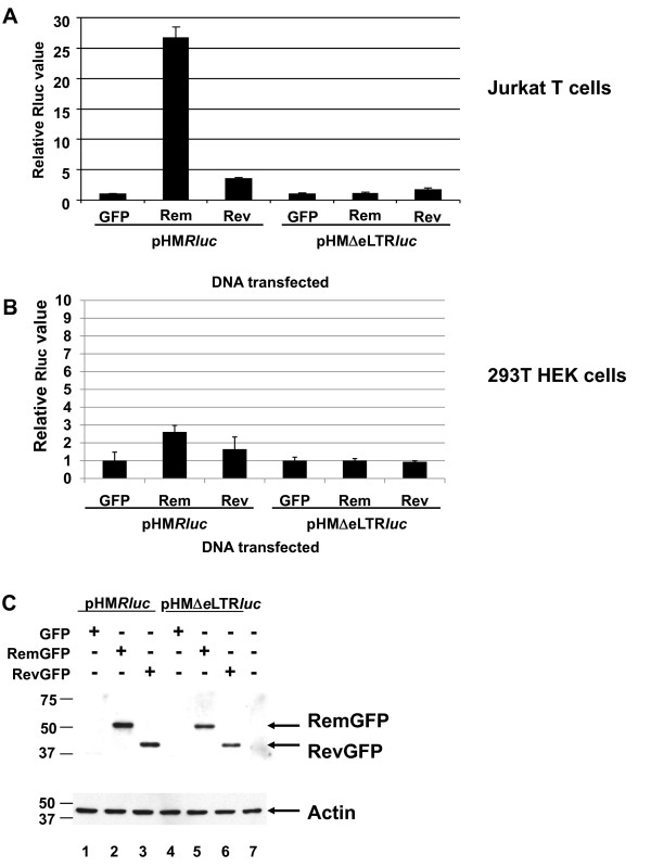
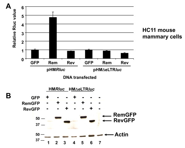
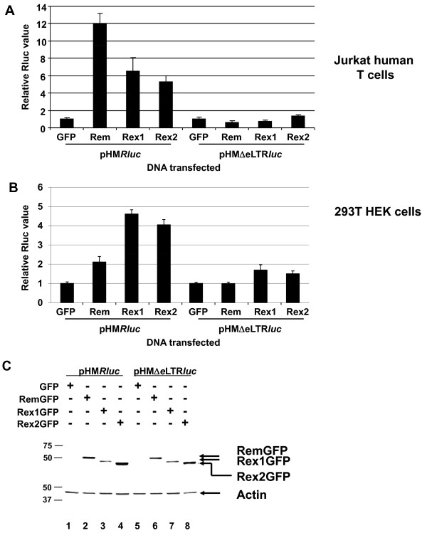
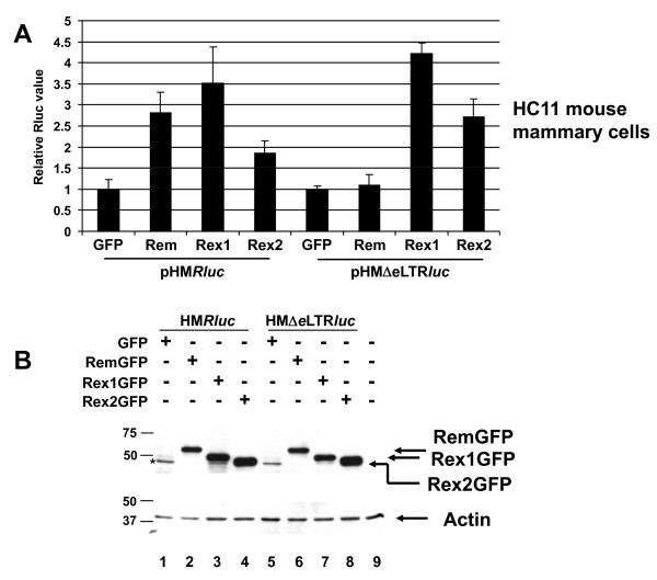
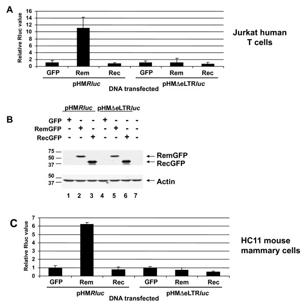
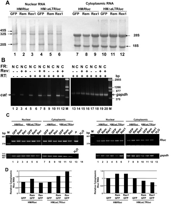
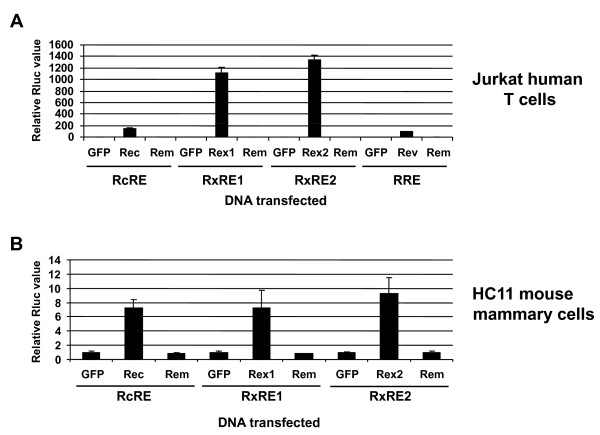
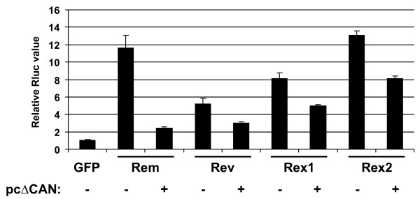
Results: MMTV Rem, HIV Rev, and HTLV Rex proteins, but not HERV-K Rec, enhanced expression from an MMTV-based reporter plasmid in human T cells, and this activity was dependent on the RmRE. No RmRE-dependent reporter gene expression was detectable using Rev, Rex, or Rec in HC11 mouse mammary cells. Cell fractionation and RNA quantitation experiments suggested that the regulatory proteins did not affect RNA stability or nuclear export in the MMTV reporter system. Rem had no demonstrable activity on export elements from HIV, HTLV, or HERV-K. Similar to the Rem-specific activity in rodent cells, the RmRE-dependent functions of Rem, Rev, or Rex in human cells were inhibited by a dominant-negative truncated nucleoporin that acts in the Crm1 pathway of RNA and protein export.

Conclusion: These data argue that many retroviral regulatory proteins recognize similar complex RNA structures, which may depend on the presence of cell-type specific proteins. Retroviral protein activity on the RmRE appears to affect a post-export function of the reporter RNA. Our results provide additional evidence that MMTV is a complex retrovirus with the potential for viral interactions in human cells.


Figures

Figure 1
Figure 1
Structure of plasmids used to determine RNA export activity. The CMV promoter (gray box) is shown inserted upstream of the 3' end of the MMTV provirus (solid horizontal line). The 3' MMTV LTR is shown by a white box. The Renilla luciferase gene (black box) is located between the splice donor (SD) and acceptor (SA) sites. The smaller hatched box indicates the SV40 polyadenylation region. The smaller gray box shows the position for insertion of response elements for other retroviral export proteins within the HMΔeLTRluc plasmid.
Figure 2
Figure 2
Activity of HIV-1 Rev on the MMTV RmRE in human cells. A. Reporter activity of RevGFP on the RmRE in Jurkat T cells. Cells were electroporated with pHMRluc or pHMΔeLTRluc (1 μg) with either 20 μg of EGFP, RemGFP or RevGFP expression plasmids. Cytoplasmic extracts were prepared and analyzed for Renilla luciferase (Rluc) activity. Average luciferase values for each reporter plasmid in the absence of Rev or Rem have been assigned a value of 1, and the other samples are reported relative to this value after normalization for DNA uptake using a co-transfected pGL3 reporter plasmid expressing firefly luciferase. Standard deviations from the average of triplicate transfections are indicated. B. Reporter activity of RevGFP on the RmRE in 293T cells. Cells were transfected using calcium phosphate precipitation of DNA as described in the Methods section. Values are reported as described in panel A. C. Western blotting confirms similar expression of Rev and Rem. A Western blot of protein extracts from Jurkat cells is shown. The unfused GFP band is not visible in this portion of the gel. The upper panel shows reactivity with GFP-specific antibody; the lower panel shows equal loading of protein extracts using an actin-specific antibody. Size markers are given in kilodaltons.
Figure 3
Figure 3
HIV-1 Rev activity on the MMTV RmRE in mouse mammary cells. A. Reporter activity in HC11 mouse mammary cells. Values are reported as described in Figure 2. B. Western blot of Rem and Rev expression in HC11 cells. Similar expression of the GFP-fusion proteins was observed as determined using antibodies specific for GFP (upper panel) or actin (lower panel). Size markers are given in kilodaltons.
Figure 4
Figure 4
Activity of HTLV Rex1 and Rex2 on MMTV RmRE-containing reporter plasmids in human cells. A. Reporter activity in Jurkat T cells. B. Reporter activity in 293T cells. Values are reported as in Figure 2, except that Rex1GFP or Rex2GFP expression plasmids were used. C. Western blotting confirms similar expression of Rem and Rex. Samples from Jurkat transfections are shown and analyzed with antibodies specific for GFP (upper panel) or actin (lower panel). Size markers are given in kilodaltons.
Figure 5
Figure 5
HTLV Rex1 and 2 activity on the MMTV RmRE in mouse mammary cells. A. Reporter activity in HC11 mouse mammary cells. Values are reported as in Figure 2, except that Rex1GFP and Rex2GFP expression plasmids were used. B. Western blots of extracts from Rex and Rem-transfected HC11 cells. A GFP-related band is observed in this blot (asterisk; lanes 1 and 5), but the major band is not visible in this portion of the gel (upper panel). Similar levels of Rem, Rex1 and Rex2 fusion proteins are observed using the GFP-specific antibody. Incubation with an actin-specific antibody revealed similar protein loading in each lane (lower panel). Size markers are given in kilodaltons.
Figure 6
Figure 6
Activity of HERV-K Rec on a reporter plasmid containing the MMTV RmRE in human and mouse cells. A. Reporter activity in Jurkat T cells. B. Western blotting of Rem and Rec expression in Jurkat cells. Results using antibodies specific for GFP (upper panel) and actin (lower panel) are shown. Size markers are given in kilodaltons. C. Reporter activity in HC11 mouse mammary cells. Values in panels A and C are reported as in Figure 2, except that a RecGFP expression plasmid was used.
Figure 7
Figure 7
Fractionation experiments indicate that Rev and Rex have little effect on export or stability of unspliced RmRE-containing reporter transcripts. A. Integrity of cytoplasmic and nuclear fractions obtained from transfected Jurkat cells. Jurkat cells were subjected to transfection by electroporation and, after 48 hr, cells were fractionated. Fractions were used for RNA extraction and subjected to Northern blotting prior to staining with methylene blue and photography. The nuclear ribosomal precursors (arrows on the left) and cytoplasmic mature ribosomal RNAs (arrows on the right) are indicated. B. Semi-quantitative RT-PCRs of fractionated RNA obtained from Jurkat cells transfected with the HIV-based reporter vector pDM128. Cells were co-transfected with pDM128 and either pEGFPN3 control vector (no Rev) or RevGFP (Rev) expression plasmids as indicated by minus or plus signs. After 48 hr, cells were fractionated, and RNA samples were extracted and subjected to RT-PCRs using primers for the cat gene or glyceraldehyde-3-phosphate dehydrogenase (gapdh). Fractions (FR) used for RNA extraction are indicated as nuclear (N) or cytoplasmic (C). PCRs performed in the absence of reverse transcriptase (RT) are indicated (lanes 1–4) (equivalent to 2 μl of a diluted cDNA reaction). Either 2 μl (lanes 5–8 and 13–16) or 4 μl (lanes 9–12 and 17–20) of the diluted cDNAs were used for RT-PCRs as indicated in Methods to show that the reactions were performed in the linear range. Samples were analyzed on a 1.5% agarose gel using either 5 (gapdh) or 15 μl (cat) of the 50 μl reaction. Markers (M) are given in basepairs (bp). C. Semi-quantitative RT-PCR assays of the MMTV-based reporter plasmids in the presence or absence of RemGFP, RevGFP, and RexGFP. Semi-quantitative RT-PCRs were performed using RNA extracted from transfected Jurkat cells and primers specific for the Renilla luciferase (Rluc) or gapdh genes. Left and right panels show results of two different transfection experiments. M = DNA markers (in bp); P = HMRluc plasmid positive control; H20 = PCR without added cDNA. D. Quantitation of RT-PCR results from cytoplasmic fractions of cells transfected with the MMTV-based reporter plasmid. Stained RT-PCR products from panel C were quantitated using ImageJ software and normalized for RNA amounts and integrity using gapdh expression. The normalized RNA levels obtained from each reporter plasmid (in the presence of the control EGFP expression vector only) were assigned values of 1, and the other samples have been reported relative to these values. These results are representative of at least three different transfection experiments.
Figure 8
Figure 8
Rem lacks activity on heterologous RNA export elements. A. Rem activity on heterologous export elements in Jurkat T cells. Cells were transfected with reporter plasmids containing the indicated response elements described in Figure 1 with or without expression vectors for retroviral export proteins. B. Rem activity on heterologous export elements in HC11 mouse mammary cells. Relative luciferase values in both panels are reported as described in Figure 2.
Figure 9
Figure 9
Rev and Rex use the Crm1 pathway to enhance expression of RmRE-containing RNAs. Jurkat cells were electroporated with 1 μg of pHMRluc and 1 μg of pGL3 control firefly luciferase plasmid. The EGFP, RemGFP, RevGFP, Rex1GFP or Rex2GFP expression plasmids (10 μg) were added as indicated with or without 20 μg of the plasmid expressing the dominant-negative nucleoporin (pcΔCAN) [42]. All samples were adjusted to the same concentration of DNA with empty vector (pBC12/CMV) prior to transfection. Luciferase activity was determined as described in Figure 2. Renilla luciferase values were normalized for DNA uptake using firefly luciferase activity, and the pHMRluc sample cotransfected with EGFP was assigned a relative value of 1.

Similar articles

Cited by

References

    1. Choi Y, Kappler JW, Marrack P. A superantigen encoded in the open reading frame of the 3' long terminal repeat of mouse mammary tumour virus. Nature. 1991;350:203–207. doi: 10.1038/350203a0. - DOI - PubMed
    1. Frankel WN, Rudy C, Coffin JM, Huber BT. Linkage of Mls genes to endogenous mammary tumour viruses of inbred mice. Nature. 1991;349:526–528. doi: 10.1038/349526a0. - DOI - PubMed
    1. Woodland DL, Lund FE, Happ MP, Blackman MA, Palmer E, Corley RB. Endogenous superantigen expression is controlled by mouse mammary tumor proviral loci. J Exp Med. 1991;174:1255–1258. doi: 10.1084/jem.174.5.1255. - DOI - PMC - PubMed
    1. Bergman AC, Bjornberg O, Nord J, Nyman PO, Rosengren AM. The protein p30, encoded at the gag-pro junction of mouse mammary tumor virus, is a dUTPase fused with a nucleocapsid protein. Virology. 1994;204:420–424. doi: 10.1006/viro.1994.1547. - DOI - PubMed
    1. Mertz JA, Simper MS, Lozano MM, Payne SM, Dudley JP. Mouse mammary tumor virus encodes a self-regulatory RNA export protein and is a complex retrovirus. J Virol. 2005;79:14737–14747. doi: 10.1128/JVI.79.23.14737-14747.2005. - DOI - PMC - PubMed

Publication types

Substances

LinkOut - more resources