. 2009 Mar 15;81(6):2106-14.
doi: 10.1021/ac802181j.

Fiber-optic microsphere-based antibody array for the analysis of inflammatory cytokines in saliva

Affiliations

Fiber-optic microsphere-based antibody array for the analysis of inflammatory cytokines in saliva

Timothy M Blicharz et al. Anal Chem. .

Abstract

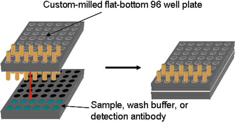
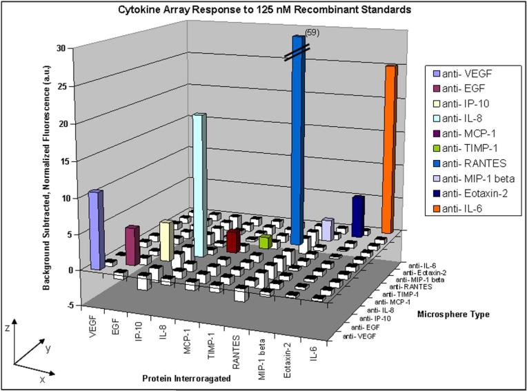
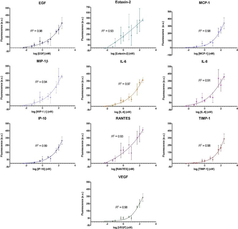
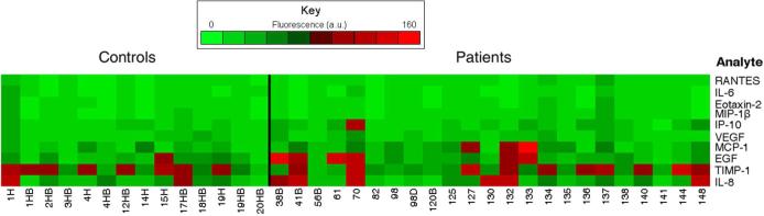
Antibody microarrays have emerged as useful tools for high-throughput protein analysis and candidate biomarker screening. We describe here the development of a multiplexed microsphere-based antibody array capable of simultaneously measuring 10 inflammatory protein mediators. Cytokine-capture microspheres were fabricated by covalently coupling monoclonal antibodies specific for cytokines of interest to fluorescently encoded 3.1 microm polymer microspheres. An optical fiber bundle containing approximately 50,000 individual 3.1 microm diameter fibers was chemically etched to create microwells in which cytokine-capture microspheres could be deposited. Microspheres were randomly distributed in the wells to produce an antibody array for performing a multiplexed sandwich immunoassay. The array responded specifically to recombinant cytokine solutions in a concentration-dependent fashion. The array was also used to examine endogenous mediator patterns in saliva supernatants from patients with pulmonary inflammatory diseases such as asthma and chronic obstructive pulmonary disease (COPD). This array technology may prove useful as a laboratory-based platform for inflammatory disease research and diagnostics, and its small footprint could also enable integration into a microfluidic cassette for use in point-of-care testing.


Figures

Figure 1
Figure 1
Principle of the fiber-optic microsphere-based antibody array. a) Capture antibodies are linked to fluorescently encoded, amine-functionalized microspheres using glutaraldehyde chemistry to produce cytokine-capture microspheres. A mixture of microspheres with different specificities is loaded into the wells of a high-density fiber-optic array to create an antibody array. b) When the array is incubated in a sample, the cytokine-capture microspheres bind their respective target analytes. The array is then incubated with a mixture of biotinylated detection antibodies. Finally, the detection antibodies are labeled with streptavidin-AF488. Note that the different microsphere types are illustrated by different colors, when they are actually encoded with different amounts of one or two fluorescent dyes.
Figure 2
Figure 2
Illustration depicting the experimental setup for parallel fiber array incubation. The wells of a microtiter plate were milled such that multiple fiber arrays could be mounted for simultaneous incubation in separate sample solutions, while being exposed to identical experimental conditions.
Figure 3
Figure 3
a) AMC registration image acquired with a 500 ms exposure to distinguish Set A and Set B microspheres (Set A microspheres are identified by colored squares, Set B microspheres are circled). b) Eu-dye registration image acquired with a 100 ms exposure to distinguish the six Set A encoding levels (Highest fluorescence intensity = blue, second highest = white, third highest = red, fourth highest = green, fifth highest = cyan, sixth highest = yellow). c) Eu-dye registration image acquired with a 500 ms exposure to distinguish the four Set B encoding levels (Highest fluorescence intensity = blue, second highest = white, third highest = red, fourth highest = cyan).
Figure 4
Figure 4
Cross-reactivity testing results using single recombinant cytokine solutions and a detection antibody cocktail. Solutions of each cytokine (125 nM) were tested using separate arrays containing all ten microsphere types (x-axis), followed by incubation with a detection antibody cocktail containing complementary antibodies for all ten cytokines. Each data point is the average fluorescence of 15−30 microspheres of each type. The net responses for all microsphere types (y-axis) were determined and normalized to three times the standard deviation of the background signals (z-axis). Responses greater than zero, shown as colored bars, indicate a positive response from a microsphere type. Responses less than zero are shown as white bars.
Figure 5
Figure 5
Standard curve analysis for each capture microsphere type on the multiplexed cytokine array.
Figure 6
Figure 6
Fiber-optic microsphere array analyses for saliva samples from healthy control individuals (left) and patients with asthma or COPD (right). While the minimum fluorescence responses for each analyte were similar between the patient and control samples, the maximum responses for several of the analytes differed substantially between the two groups. The responses were as follows: IP-10 (patient = 83, control = 26), MCP-1 (patient = 144, control = 52), and EGF (patient = 127, control = 68) (Fluorescence reported as arbitrary units).
Figure 7
Figure 7
Mean microsphere responses for pulmonary inflammatory disease patients (red) and control samples (blue) tested with microsphere-based antibody arrays. Greater variability was observed in pulmonary patient samples for several of the biomarkers (IL-8, TIMP-1, EGF, MCP-1, and IP-10), as is illustrated by larger standard deviations.

References

    1. Urbanowska T, Mangialaio S, Zickler C, Cheevapruk S, Hasler P, Regenass S, Legay F. J. Immunol. Methods. 2006;316:1–7. - PubMed
    1. Angenendt P. Drug Discov. Today. 2005;10:503–511. - PubMed
    1. Gao W-M, Kuick R, Orchekowski RP, Misek DE, Qiu J, Greenberg AK, Rom WN, Brenner DE, Omenn GS, Haab BB, Hanash SM. BMC Cancer. 2005;5 No pp. given.
    1. Nielsen UB, Geierstanger BH. J. Immunol. Methods. 2004;290:107–120. - PubMed
    1. Holgate ST, Polosa R. Nat. Rev. Immunol. 2008;8:218–230. - PubMed

Publication types

MeSH terms