

A rare coexistence of adrenal cavernous hemangioma with extramedullar hemopoietic tissue: a case report and brief review of the literature
- PMID: 19193247
- PMCID: PMC2647540
- DOI: 10.1186/1477-7819-7-13
A rare coexistence of adrenal cavernous hemangioma with extramedullar hemopoietic tissue: a case report and brief review of the literature
Abstract
Background: Cavernous hemangiomas of the adrenal gland are rare, benign, non-functioning neoplastic tumors. To our knowledge, 55 cases have been reported in the literature to date.
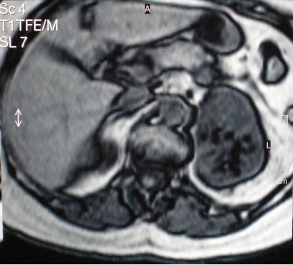
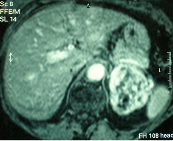
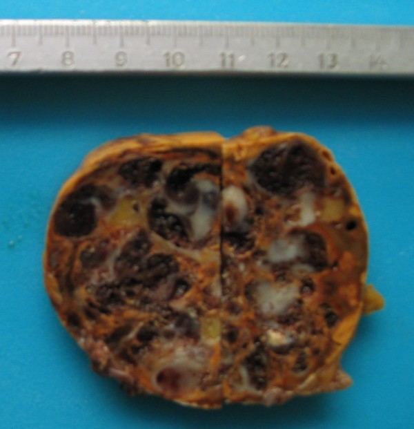
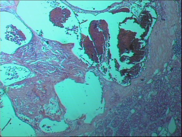
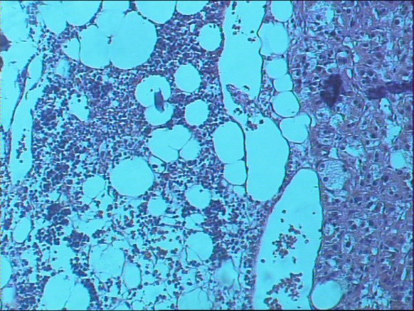
Case presentation: We report the first case of a large, non-functioning adrenal cavernous hemangioma that was incidentally found during the preoperative staging workup of a 75 year old woman with left breast adenocarcinoma. Imaging with US, CT scan and MRI showed a heterogeneous 8 cm mass with non-specific radiological features that was located on the left adrenal gland. The mass was surgically excised and pathology revealed an adrenal hemangioma with areas of extramedullar hemopoiesis.
Conclusion: Although adrenal hemangiomas are rare and their preoperative diagnosis is difficult, they should always be included in the differential diagnosis of adrenal neoplasms.
Figures
Similar articles
-
Adrenal cavernous hemangioma: a case report.BMC Surg. 2018 Nov 20;18(1):103. doi: 10.1186/s12893-018-0429-9. BMC Surg. 2018. PMID: 30458815 Free PMC article.
-
A case of adrenal cavernous hemangioma.Int J Urol. 1997 Nov;4(6):608-10. doi: 10.1111/j.1442-2042.1997.tb00318.x. Int J Urol. 1997. PMID: 9477193
-
Rare cavernous hemangioma of adrenal gland: case report.Sao Paulo Med J. 2014;132(4):249-52. doi: 10.1590/1516-3180.2014.1324715. Sao Paulo Med J. 2014. PMID: 25055072 Free PMC article.
-
Adrenal cavernous hemangioma: a case report with review of the literature.JOP. 2014 May 27;15(3):254-7. doi: 10.6092/1590-8577/2402. JOP. 2014. PMID: 24865537 Review.
-
Cavernous adrenal hemangioma.Urology. 1993 Sep;42(3):327-30. doi: 10.1016/0090-4295(93)90626-l. Urology. 1993. PMID: 8379036 Review.
Cited by
-
Incidental adrenal hemangioma clinically suspicious for malignancy: diagnostic considerations and review of the literature.Int J Clin Exp Pathol. 2022 Nov 15;15(11):444-458. eCollection 2022. Int J Clin Exp Pathol. 2022. PMID: 36507066 Free PMC article.
-
A Rare Cavernous Hemangioma of the Adrenal Gland.Urol Case Rep. 2015 Apr 29;3(4):120-2. doi: 10.1016/j.eucr.2015.03.009. eCollection 2015 Jul. Urol Case Rep. 2015. PMID: 26793524 Free PMC article.
-
The diagnostic dilemma of adrenal vascular tumors: analysis of 21 cases and systematic review of the literature.Endocrine. 2025 Mar;87(3):1291-1304. doi: 10.1007/s12020-024-04123-5. Epub 2025 Jan 18. Endocrine. 2025. PMID: 39825193 Free PMC article.
-
Adrenal cavernous hemangioma: a case report.BMC Surg. 2018 Nov 20;18(1):103. doi: 10.1186/s12893-018-0429-9. BMC Surg. 2018. PMID: 30458815 Free PMC article.
-
Adrenal Cavernous Hemangioma: A Rarely Perceived Pathology-Case Illustration and Review of Literature.Case Rep Pathol. 2019 Dec 17;2019:8463890. doi: 10.1155/2019/8463890. eCollection 2019. Case Rep Pathol. 2019. PMID: 31949968 Free PMC article.
References
-
- Johnson CC, Jeppesen FB. Haemangioma of the adrenal. J Urol. 1955;74:573–577. - PubMed
-
- Heis HA, Bani-Hani KE, Bani-Hani BK. Adrenal cavernous haemangioma. Singapore Med J. 2008;49:e236–e237. - PubMed
-
- Makiyama K, Fukuoka H, Kawamoto K, Suwa Y. Surgical removal of adrenal haemangioma after five years of follow-up: a case report. Hinyokika Kiyo. 1998;44:579–581. - PubMed
Publication types
MeSH terms
LinkOut - more resources
Full Text Sources
Medical
