. 2009 Mar 30;386(1):109-21.
doi: 10.1016/j.virol.2009.01.007. Epub 2009 Feb 3.

Co-immunization with IL-15 enhances cellular immune responses induced by a vif-deleted simian immunodeficiency virus proviral DNA vaccine and confers partial protection against vaginal challenge with SIVmac251

Affiliations

Co-immunization with IL-15 enhances cellular immune responses induced by a vif-deleted simian immunodeficiency virus proviral DNA vaccine and confers partial protection against vaginal challenge with SIVmac251

Robert A Dubie et al. Virology. .

Abstract

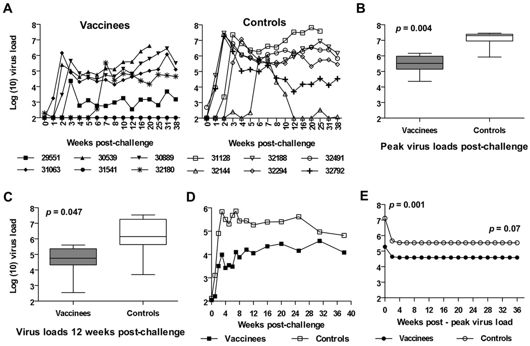
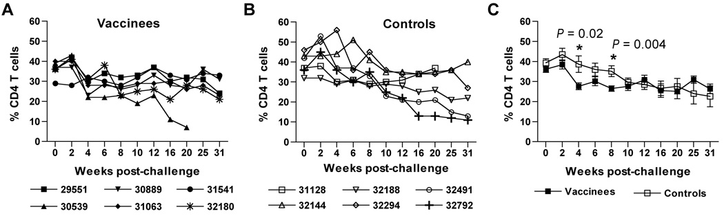
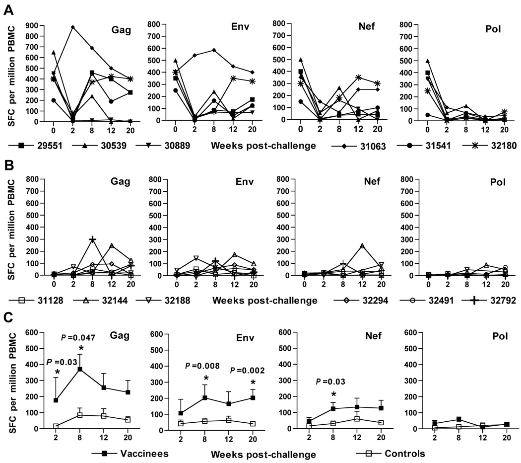
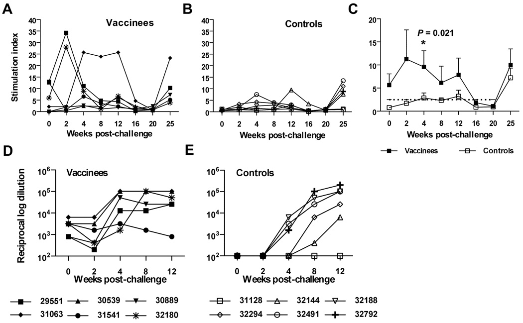
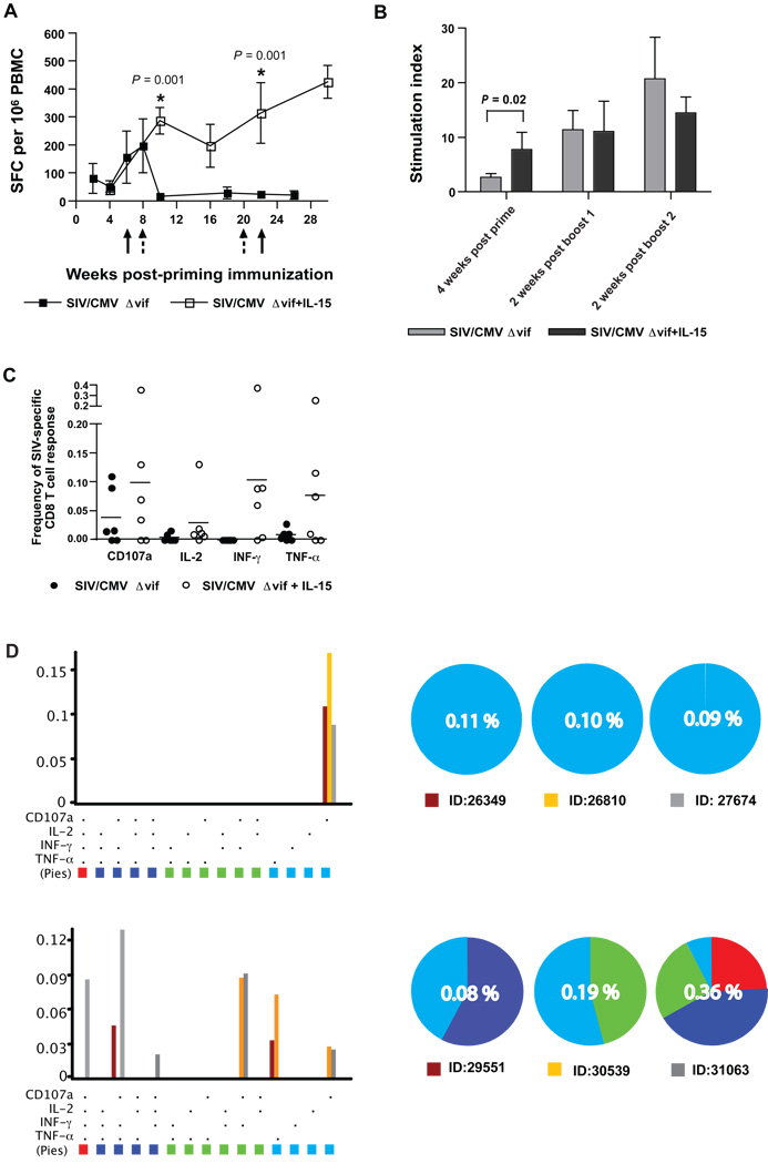
Simian immunodeficiency virus (SIV) infection of rhesus macaques is a valuable animal model for human immunodeficiency virus (HIV)-1 vaccine development. Our laboratory recently described the immunogenicity and limited efficacy of a vif-deleted SIVmac239 proviral DNA (SIV/CMVDelta vif) vaccine. The current report characterizes immunogenicity and efficacy for the SIV/CMVDelta vif proviral DNA vaccine when co-inoculated with an optimized rhesus interleukin (rIL)-15 expression plasmid. Macaques co-inoculated with rIL-15 and SIV/CMVDelta vif proviral plasmids showed significantly improved SIV-specific CD8 T cell immunity characterized by increased IFN-gamma ELISPOT and polyfunctional CD8 T cell responses. Furthermore, these animals demonstrated a sustained suppression of plasma virus loads after multiple low dose vaginal challenges with pathogenic SIVmac251. Importantly, SIV-specific cellular responses were greater in immunized animals compared to unvaccinated controls during the initial 12 weeks after challenge. Taken together, these findings support the use of IL-15 as an adjuvant in prophylactic anti-HIV vaccine strategies.


Figures

Figure 1
Figure 1. Production of rIL-15 from an optimized expression plasmid
Expression plasmids designated pNDRRm-IL-15 and pNDgmsRRm-IL-15, were constructed as described in Materials and Methods and were distinguished by their leader sequences. Cell culture supernatants were harvested at two, four, and six days from either COS-7 or 293T cells, after transfection with a rIL-15 expression plasmid and tested for IL-15 concentrations by a commercial human IL-15 antigen-capture ELISA (Invitrogen, Biosource) as described in Materials and Methods. Error bars represent standard deviation.
Figure 2
Figure 2. SIV-specific IFN-γ ELISPOT responses elicited by co-immunization with SIV/CMVΔvif and pNDgmsRRm-IL-15 plasmid DNA
SIV-specific IFN-γ ELISPOT responses were assayed on cryopreserved PBMC harvested from animals at various time points after vaccination. Immune responses to individual SIVmac239 proteins were measured using recombinant vaccinia viruses expressing either SIV Gag, Env, Nef or Pol, with protocols described in Materials and Methods. Values less than 50 spot-forming colonies (SFC) per 106 PBMC were considered nonspecific for this assay. Arrows positioned at 8 and 20 weeks after priming immunization denoted time points of booster vaccine inoculations.
Figure 3
Figure 3. SIV-specific T cell proliferative and antiviral antibody responses elicited by co-immunization with SIV/CMVΔvif plasmid DNA and pNDgmsRRm-IL-15 plasmid DNA
Virus-specific T cell proliferative responses directed to purified inactivated SIV were measured in PBMC harvested from macaques at various time points after priming immunization. Values for the highest SI to either 12.5 or 125 ng of SIV p27Gag are shown (A). A SI of 2.5 or greater was considered positive. Reciprocal endpoint titers for plasma antibodies against native SIV Env were measured using a ConA ELISA with reciprocal log dilution values greater than 100 representing positive titers (B). Arrows positioned at 8 and 20 weeks after priming immunization denoted time points of booster vaccine inoculations.
Figure 4
Figure 4. Plasma virus loads after multiple low dose IVAG challenge of vaccinated and unvaccinated macaques with SIVmac251
Plasma virus loads represented as SIV RNA copies per ml of plasma, was determined after SIVmac251 challenge by a real-time RT-PCR assay for SIV gag RNA for vaccinated and unvaccinated macaques (A). Peak virus loads (B) and virus loads measured at 12 weeks after initial challenge (C) are compared between viremic vaccinated and unvaccinated macaques using the Mann-Whitney U test with the uninfected vaccinated animal excluded from the analysis. Geometric means for plasma RNA loads for all vaccinated and control macaques over time after the initial challenge inoculation are also shown (D). In a different analysis, a nonlinear mixed-effects model based on an exponential function was fitted to log RNA values, with plasma RNA concentrations recorded from the first observation of peak plasma viral RNA, to provide mean virus load curves for vaccinated and unvaccinated macaques through the duration of the study (E), also with the uninfected vaccinated animal excluded from the analysis.
Figure 5
Figure 5. Quantitation of CD4 T cell percentages after challenge with SIVmac251
Peripheral blood CD4 T cell percentages were calculated for vaccinated (A) and unvaccinated control (B) macaques after SIVmac251 challenge. Mean values with standard error for CD4 T cell percentages over time challenge are shown for both groups of animals (C). Asterisks (*) positioned over values at 4 and 8 weeks after challenge indicate a significant difference between vaccinated and SIV-infected unvaccinated controls as determined by a pair-wise comparison using a Mann-Whitney U test.
Figure 6
Figure 6. IFN-γ ELISPOT responses in animals after challenge with SIVmac251
Virus-specific IFN-γ ELISPOT responses were measured for vaccinated (A) and unvaccinated (B) animals over time after the initial challenge inoculation. Mean values for SFC with standard error over time after challenge are shown for both groups of animals (C). Asterisks (*) positioned over values indicate a significant difference between vaccinated and SIV-infected unvaccinated controls as determined by a pair-wise comparison using a Mann-Whitney U test.
Figure 7
Figure 7. SIV-specific T cell proliferative responses and SIV Env antibody responses in animals after challenge with SIVmac251
Virus-specific T cell proliferative responses are shown for vaccinated (A) and unvaccinated control (B) animals over time after the initial challenge inoculation. Mean values for SI with standard error over time after challenge are shown for both groups of animals (C). A SI of 2.5 or greater was considered positive (see dashed cut-line). Asterisks (*) positioned over values indicate a significant difference between vaccinated and SIV-infected unvaccinated controls as determined by a pair-wise comparison using a Mann-Whitney U test. Reciprocal endpoint titers for plasma antibodies against native SIV Env are described for vaccinated (D) and unvaccinated control (E) animals over consecutive time points after challenge. Reciprocal endpoint titers > 100 were considered positive.
Figure 8
Figure 8. Adjuvant effects on SIV-specific cellular immune responses conferred by IL-15
Mean values for SIV-specific IFN-γ ELISPOT (SFC) to SIV Gag with standard error over time after priming immunization are shown for animals vaccinated with either SIV/CMVΔvif DNA alone, or with both SIV/CMVΔvif and pNDgmsRRm-IL-15 plasmid DNA (A). Arrows with a solid line denote booster immunization time points for animals vaccinated with SIV/CMVΔvif DNA alone. Arrows with a dashed line denote booster immunization time points for animals vaccinated with SIV/CMVΔvif DNA and pNDgmsRRm-IL-15 plasmids. Asterisks (*) positioned over values indicate a significant difference between values measured for the two different vaccine groups, as determined by a pair-wise comparison using a Mann-Whitney U test. Similarly, mean values for peripheral blood SIV-specific T cell proliferation responses (SI) over time after priming and booster immunizations are shown for animals vaccinated with SIV/CMVΔvif DNA alone or also with pNDgmsRRm-IL-15 plasmid (B). Frequencies of peripheral blood CD8 T cells expressing cytokines and a degranulation marker CD107a after exposure to a SIV capsid peptide pool were measured by CFC at 2 weeks after a second DNA vaccine booster immunization and compared between the two vaccine groups (C). Significant differences between frequencies for each immune response marker for the two vaccine groups were not observed using a pair-wise comparison using a Mann-Whitney U test. Frequencies of peripheral blood CD8 T cells expressing multiple immune response markers, also at 2 weeks after a second DNA vaccine booster immunization, were compared between the two vaccine groups (D). Bar graphs on the left show the percentages of SIV Gag-specific CD8 T cells in each of 15 categories representing all possible combinations of the cytokines and the degranulation marker analyzed. Cells positive for all four markers (i.e., IL-2, IFN-γ, TNF-α and CD107) are indicated in red. Cells positive for three markers are shown in dark blue; those positive for two functions in green; and those positive for a single function in light blue. Pie charts convert the total capsid-specific CD8 T-cell response in each macaque to 100%, with 4-, 3-, 2-, and 1-function cells shown as individual slices of the pie. Pie slices are color-coded to match the bar graphs. Numbers in the center of each pie indicate the total capsid-specific CD8 T-cell response, with all functional categories combined. ID denotes animal number and bar color for that particular animal represented on the graph.

References

    1. Abel K, Compton L, Rourke T, Montefiori D, Lu D, Rothaeusler K, Fritts L, Bost K, Miller CJ. Simian-human immunodeficiency virus SHIV89.6-induced protection against intravaginal challenge with pathogenic SIVmac239 is independent of the route of immunization and is associated with a combination of cytotoxic T-lymphocyte and alpha interferon responses. J. Virol. 2003;77:3099–3118. - PMC - PubMed
    1. Almeida JR, Price DA, Papagno L, Arkoub ZA, Sauce D, Bornstein E, Asher TE, Samri A, Schnuriger A, Theodorou I, Costagliola D, Rouzioux C, Agut H, Marcelin AG, Douek D, Autran B, Appay V. Superior control of HIV-1 replication by CD8+ T cells is reflected by their avidity, polyfunctionality, and clonal turnover. J. Exp. Med. 2007;204:2473–2485. - PMC - PubMed
    1. Becker TC, Wherry EJ, Boone D, Murali-Krishna K, Antia R, Ma A, Ahmed R. Interleukin 15 is required for proliferative renewal of virus-specific memory CD8 T cells. J. Exp. Med. 2002;195:1541–1542. - PMC - PubMed
    1. Boyer JD, Robinson TM, Kutzler MA, Vansant G, Hokey DA, Kumar S, Parkinson R, Wu L, Sidhu MK, Pavlakis GN, Felber BK, Brown C, Silvera P, Lewis MG, Monforte J, Waldmann TA, Eldridge J, Weiner DB. Protection against simian/human immunodeficiency virus (SHIV) 89.6P in macaques after coimmunization with SHIV antigen and IL-15 plasmid. Proc. Natl. Acad. Sci. USA. 2007;104:18648–18653. - PMC - PubMed
    1. Calarota SA, Weiner DB. Approaches for the design and evaluation of HIV-1 DNA vaccines. Expert Rev. Vaccines. 2004;3:S135–S149. - PubMed

Publication types

MeSH terms