. 2009 Jun;8(6):1424-35.
doi: 10.1074/mcp.M800571-MCP200. Epub 2009 Feb 3.

Functional and complementary phosphorylation state attributes of human insulin-like growth factor-binding protein-1 (IGFBP-1) isoforms resolved by free flow electrophoresis

Affiliations

Functional and complementary phosphorylation state attributes of human insulin-like growth factor-binding protein-1 (IGFBP-1) isoforms resolved by free flow electrophoresis

Mikkel Nissum et al. Mol Cell Proteomics. 2009 Jun.

Abstract

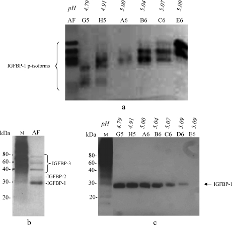
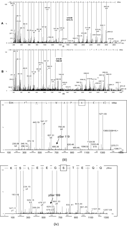
Fetal growth restriction (FGR) is a common disorder in which a fetus is unable to achieve its genetically determined potential size. High concentrations of insulin-like growth factor-binding protein-1 (IGFBP-1) have been associated with FGR. Phosphorylation of IGFBP-1 is a mechanism by which insulin-like growth factor-I (IGF-I) bioavailability can be modulated in FGR. In this study a novel strategy was designed to determine a link between IGF-I affinity and the concomitant phosphorylation state characteristics of IGFBP-1 phosphoisoforms. Using free flow electrophoresis (FFE), multiple IGFBP-1 phosphoisoforms in amniotic fluid were resolved within pH 4.43-5.09. The binding of IGFBP-1 for IGF-I in each FFE fraction was determined with BIAcore biosensor analysis. The IGF-I affinity (K(D)) for different IGFBP-1 isoforms ranged between 1.12e-08 and 4.59e-07. LC-MS/MS characterization revealed four phosphorylation sites, Ser(P)(98), Ser(P)(101), Ser(P)(119), and Ser(P)(169), of which Ser(P)(98) was new. Although the IGF-I binding affinity for IGFBP-1 phosphoisoforms across the FFE fractions did not correlate with phosphopeptide intensities for Ser(P)(101), Ser(P)(98), and Ser(P)(169) sites, a clear association was recorded with Ser(P)(119). Our data demonstrate that phosphorylation at Ser(119) plays a significant role in modulating affinity of IGFBP-1 for IGF-I. In addition, an altered profile of IGFBP-1 phosphoisoforms was revealed between FGR and healthy pregnancies that may result from potential site-specific phosphorylation. This study provides a strong basis for use of this novel approach in establishing the linkage between phosphorylation of IGFBP-1 and FGR. This overall strategy will also be broadly applicable to other phosphoproteins with clinical and functional significance.


Figures

F<sc>ig</sc>. 1.
Fig. 1.
Enrichment and detection of IGFBP-1 in AF. a, SDS gel (12% PAGE) with silver staining. Lane 1, crude AF; Lane 2, enriched AF (total protein, 28 μg/lane). An enrichment of IGFBP-1 (∼30 kDa) was achieved with a reduction in band (∼60 kDa) intensity for albumin (Lane 2). b, immunoblot analysis using anti-IGFBP-1 mAb 6303. Lane 1, crude AF; Lane 2, enriched AF (total protein, 8 μg) showing intact IGFBP-1 and its proteolytic fragment. c–e, 2-D immunoblot analysis using anti IGFBP-1 polyclonal antibody. c, crude AF; d, enriched AF (∼25 μg of total protein) with multiple IGFBP-1 phosphoisoforms represented by the numbered spots in each panel. e, AF sample following alkaline phosphatase treatment with dephosphorylated IGFBP-1. Lane M, molecular mass markers.
F<sc>ig</sc>. 2.
Fig. 2.
a, FFE pH gradients used for the separation of AF proteins. The flat regions below pH 2 and above pH 12 in the broad range pH gradient (DSE) (i) and below pH 3 and above pH 7 in the narrow range pH gradient (ii) represent the pH values of the anodic and cathodic stabilization media, respectively. Fractions (1–96) and their positions in the sample collection plates (A1–H12) are indicated. Sample preparation and FFE fractionation were performed as described in the text. The range of FFE fractions (C5–B7) used for the analysis of IGFBP-1 from the DSE separation is also indicated. b, SDS-PAGE with SilverQuest staining of FFE-fractionated AF. The distribution of total proteins is shown in fractions A4 to C10 (pH 1.6–12.4). FFE samples (10 μl/lane) were loaded. Lane M is the molecular mass marker, and Lane S is an aliquot of the enriched AF used in FFE separation. The perspective range of fractions for analysis of IGFBP-1 isoforms is indicated. c, immunoblot analysis of FFE fractions using mAb 6303. Lane M is the molecular mass marker, and E5 to D6 FFE fractions (20 μl/lane) (DSE separation) are indicated with respective pH values.
F<sc>ig</sc>. 3.
Fig. 3.
IGFBP-1 phosphoisoform separation using native gel electrophoresis. a, immunoblot analysis using mAb 6303 combined with native gel analysis. Lane AF, crude AF (∼50 ng of total IGFBP-1); Lanes G5E6, equal volumes (30 μl/lane) of the FFE fractions loaded showing multiple bands. In fractions G5 to E6 variable intensities of the double bands indicate low to high abundance of IGFBP-1 phosphoisoforms (p-isoforms) that are partially overlapping in the individual fractions. b and c, ligand blot analysis using biotinylated rIGF-I showing crude AF (b, Lane AF; 5 μl) and individual FFE fractions (c, Lanes G5–E6; 5 μl). Several IGF-I-binding proteins were identified in AF by their molecular mass, whereas only IGFBP-1 was detected in the FFE fractions. Lane M, molecular mass markers.
F<sc>ig</sc>. 4.
Fig. 4.
BIAcore biosensor interaction of immobilized rIGF-I with IGFBP-1 displaying the association and dissociation phases of concentration-dependent IGFBP-1 binding with rIGF-I. a–c, IGFBP-1 phosphoisoform(s) present in FFE fractions E5 (pH 4.43) and F6 (pH 5.09) and in the enriched AF, respectively. The kinetics analysis shows differences between dissociation phases for the phosphoisoforms separated at the lower pH (4.91) compared with the higher pH (5.09) or the unfractionated AF.
F<sc>ig</sc>. 5.
Fig. 5.
Representative LC-MS/MS ion spectra of IGFBP-1 phosphopeptides identified in FFE fraction C6. The peptide sequences with highlighted IGFBP-1 phosphorylation sites are also indicated for each spectrum. Shown is the deconvoluted spectra of the parent ion at m/z 949.73, representing the singly phosphorylated Ser101 (Ser(P)101 with the peptide sequence in (i) and m/z 976.42 representing the doubly phosphorylated peptide (Ser(P)101 and Ser(P)98) in (ii). Both ions were observed as triply charged ions. In spectrum (i), intense b ions confirm the amino acid sequence of the peptide. The observed b18 ion at m/z 1771.68 and the b18 − 98 ion at m/z 1673.75 that is derived from b18 ion with a loss of H3PO4 confirmed phosphorylation on the Ser101 residue. In spectrum (ii), the precursor ion is 80 Da heavier than the ion in spectrum (i), indicating additional phosphorylation. The observed b15 ion at m/z 1458.63 and the b15 − 98 ion at m/z 1360.59 confirmed phosphorylation on Ser98 in addition to Ser101. Also are shown representative LC-MS/MS ion spectra of IGFBP-1 phosphopeptides identified with Ser(P)119, 685.30 m/z, charge state +2 (iii) and Ser(P)169, m/z 629.78, charge state +2 in (iv) using an aliquot of FFE sample C6.
F<sc>ig</sc>. 6.
Fig. 6.
2-D immunoblot analysis of IGFBP-1 phosphoisoforms in crude AF. Samples (total protein, 25 μg/AF sample) were analyzed using anti-IGFBP-1 polyclonal antibody. a, normal (healthy) (n = 6); b, FGR pregnancies (n = 6). The patterns of spots representing multiple IGFBP-1 phosphoisoforms were compared visually.

Similar articles

Cited by

References

    1. Martina, N. A., Kim, E., Chitkara, U., Wathen, N. C., Chard, T., and Giudice, L. C. ( 1997) Gestational age-dependent expression of insulin-like growth factor-binding protein-1 (IGFBP-1) phosphoisoforms in human extraembryonic cavities, maternal serum, and decidua suggests decidua as the primary source of IGFBP-1 in these fluids during early pregnancy. J. Clin. Endocrinol. Metab. 82, 1894–1898 - PubMed
    1. Rajaram, S., Baylink, D. J., and Mohan, S. ( 1997) Insulin-like growth factor-binding proteins in serum and other biological fluids: Regulation and functions. Endocr. Rev. 18, 801–831 - PubMed
    1. Coverley, J. A., and Baxter, R. C. ( 1997) Phosphorylation of insulin-like growth factor binding proteins. Mol. Cell. Endocrinol. 128, 1–5 - PubMed
    1. Gibson, J. M., Aplin, J. D., White, A., and Westwood, M. ( 2001) Regulation of IGF bioavailability in pregnancy. Mol. Hum. Reprod. 7, 79–87 - PubMed
    1. Yu, J., Iwashita, M., Kudo, Y., and Takeda, Y. ( 1998) Phosphorylated insulin-like growth factor (IGF)-binding protein-1 (IGFBP-1) inhibits while non-phosphorylated IGFBP-1 stimulates IGF-I-induced amino acid uptake by cultured trophoblast cells. Growth Horm. IGF Res. 8, 65–70 - PubMed

Publication types

MeSH terms

Substances

LinkOut - more resources