. 2009 May;8(5):1150-64.
doi: 10.1074/mcp.M800193-MCP200. Epub 2009 Feb 3.

Affinity purification strategy to capture human endogenous proteasome complexes diversity and to identify proteasome-interacting proteins

Affiliations

Affinity purification strategy to capture human endogenous proteasome complexes diversity and to identify proteasome-interacting proteins

Marie-Pierre Bousquet-Dubouch et al. Mol Cell Proteomics. 2009 May.

Abstract

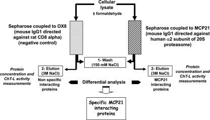
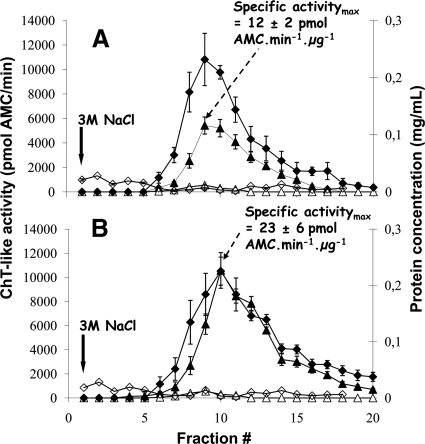
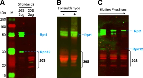
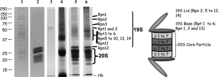
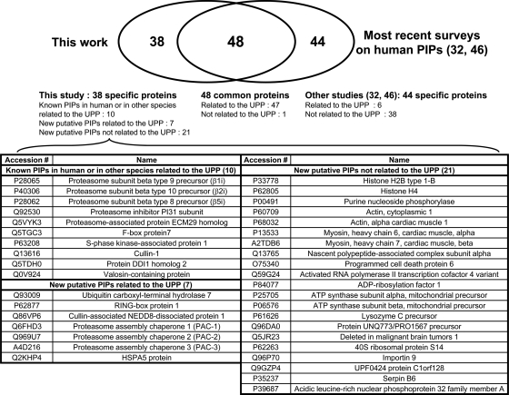
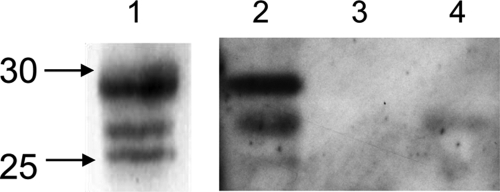
An affinity purification strategy was developed to characterize human proteasome complexes diversity as well as endogenous proteasome-interacting proteins (PIPs). This single step procedure, initially used for 20 S proteasome purification, was adapted to purify all existing physiological proteasome complexes associated to their various regulatory complexes and to their interacting partners. The method was applied to the purification of proteasome complexes and their PIPs from human erythrocytes but can be used to purify proteasomes from any human sample as starting material. The benefit of in vivo formaldehyde cross-linking as a stabilizer of protein-protein interactions was studied by comparing the status of purified proteasomes and the identified proteins in both protocols (with or without formaldehyde cross-linking). Subsequent proteomics analyses identified all proteasomal subunits, known regulators, and recently assigned partners. Moreover other proteins implicated at different levels of the ubiquitin-proteasome system were also identified for the first time as PIPs. One of them, the ubiquitin-specific protease USP7, also known as HAUSP, is an important player in the p53-HDM2 pathway. The specificity of the interaction was further confirmed using a complementary approach that consisted of the reverse immunoprecipitation with HAUSP as a bait. Altogether we provide a valuable tool that should contribute, through the identification of partners likely to affect proteasomal function, to a better understanding of this complex proteolytic machinery in any living human cell and/or organ/tissue and in different cell physiological states.


Figures

F<sc>ig</sc>. 1.
Fig. 1.
Differential analysis strategy to identify specific human PIPs. Erythrocytes submitted or not to in vivo cross-linking were lysed, and proteins were purified by overnight incubation at 4 °C with either the MCP21-Sepharose antibody or the OX8-Sepharose antibody as negative control. After extensive washing with 20 mm Tris-HCl, 150 mm NaCl, 1 mm EDTA, 10% glycerol, 5 mm MgCl2, 2 mm ATP, pH 7.6, proteins were eluted with a saline step using the same buffer containing 3 m NaCl. Two-milliliter fractions were collected and stored at 4 °C. Protein concentration was determined using the Bio-Rad Protein Assay, and proteasome content could be estimated by measuring the in vitro chymotrypsin-like activity as described under “Experimental Procedures.” The fractions containing the most proteasome activity were then further analyzed by Western blotting and by SDS-PAGE followed by trypsin digestion and LC-MS/MS analysis. Proteins identified in each case (experiment and negative control) were subjected to a differential analysis using the MFPaQ software (38) so that a list of specific MCP21-interacting proteins could be generated. ChT-L, ChT-like.
F<sc>ig</sc>. 2.
Fig. 2.
Proteasome purification from human erythrocytes without (A) or with (B) in vivo formaldehyde cross-linking. Proteasome complexes were purified from human erythrocytes by affinity chromatography with the mouse IgG1 monoclonal antibody MCP21 coupled to Sepharose beads. A control purification was performed using the mouse IgG1 monoclonal antibody OX8 directed against rat CD8α. Proteins were cross-linked in vivo by incubation of human erythrocytes with 1% formaldehyde as indicated under “Experimental Procedures.” After incubation of 50 ml of erythrocyte lysate with either the MCP21-Sepharose or the OX8-Sepharose, proteins interacting with the beads were eluted by a saline step of 3 m NaCl. The eluted fractions were analyzed for their protein concentrations as well as for their proteasome contents. Statistical results were obtained from three independent experiments for each condition (formaldehyde-treated or not). Error bars indicate standard deviations (n = 3). ♦ and ⋄ represent protein concentrations in the fractions from the MCP21-Sepharose beads and from the OX8-Sepharose beads, respectively. ▴ and ▵ represent the in vitro ChT-like activity without and with 10 μm lactacystin, respectively, in the fractions eluted from the MCP21-Sepharose beads. No ChT-like activity could be detected in the fractions from the OX8 negative control experiment.
F<sc>ig</sc>. 3.
Fig. 3.
Detection of 20 S core particle and 19 S activators in the purified proteasome preparations. Proteins separated by SDS-PAGE were transferred to a nitrocellulose membrane: 2 μg of commercial 20 S and 26 S proteasome from human erythrocytes as standards (A), 20 μg of total proteins from fractions containing the maximal ChT-like activity (fractions 9 and 10 from purifications without and with formaldehyde cross-linking, respectively) (B), and 10 μg of estimated 20 S proteasome (based on the ChT-like activity measurement) from fractions 8, 9, and 10 from the purification without formaldehyde cross-linking (C). Mouse monoclonal primary antibodies against 19 S proteasome subunits Rpt1 and Rpn12 and rabbit polyclonal antibodies against 20 S core subunits were used for the immunoblot staining. ECL Plex CyDye-conjugated antibodies, goat α-mouse IgG-Cy3 and goat α-rabbit IgG-Cy5, were used as secondary antibodies. The detection was performed using the Typhoon Trio fluorescence scanner at 532 nm excitation and 580 nm emission for the Cy3-conjugated antibody and 633 nm excitation and 670 nm emission for the Cy5-conjugated antibody. Lane M, molecular mass markers.
F<sc>ig</sc>. 4.
Fig. 4.
Separation of proteasomes and proteasome-interacting proteins by SDS-PAGE. Proteins were separated by SDS-PAGE on a 8 × 10-cm acrylamide gel (12%) and detected by Coomassie Brilliant Blue staining. Whole gel lanes were analyzed. In-gel tryptic digestion of proteins was performed prior to identification by nano-LC-ESI-LTQ-Orbitrap MS/MS analysis and database searching as described under “Experimental Procedures.” Lanes 1 and 2, 2 μg of commercially available human erythrocyte 26 S and 20 S proteasomes, respectively; lanes 3 and 4, the whole protein content of eluted proteins from the OX8-Sepharose was loaded after concentration by ultrafiltration (lane 3, non formaldehyde-treated erythrocytes; lane 4, formaldehyde-treated erythrocytes); lanes 5 and 6, 40 μg of proteins from fractions 6–9 and 7–10 from purifications without and with formaldehyde cross-linking, respectively.
F<sc>ig</sc>. 5.
Fig. 5.
Venn diagram illustrating the overlap between human PIPs identified by us and the most recent surveys on human PIPs interaction data obtained by TAP strategies followed by systematic MS identification of proteins (32, 46).
F<sc>ig</sc>. 6.
Fig. 6.
Detection of 20 S core particle in the immunoprecipitated HAUSP complexes. HAUSP complexes were immunoprecipitated using the anti-HAUSP antibody or a control antibody as described under “Experimental Procedures,” separated by SDS-PAGE, and transferred to a nitrocellulose membrane. Rabbit polyclonal antibodies against 20 S core subunits were used for the immunoblot staining. Lane 1, 0.05 μg of commercially available human erythrocyte 20 S proteasome; lanes 2 and 4, whole protein content of eluted proteins from a 1.5-ml erythrocyte aliquot precipitated with the anti-HAUSP antibody (lane 2) or with the OX8 antibody (control) (lane 4); lane 3, protein sample eluted from the anti-HAUSP-Dynabeads after incubation with 1.5 ml of lysis buffer.

References

    1. Hershko, A., and Ciechanover, A. ( 1998) The ubiquitin system. Annu. Rev. Biochem. 67, 425–479 - PubMed
    1. Glickman, M. H., and Ciechanover, A. ( 2002) The ubiquitin-proteasome proteolytic pathway: destruction for the sake of construction. Physiol. Rev. 82, 373–428 - PubMed
    1. Ciechanover, A., Orian, A., and Schwartz, A. L. ( 2000) Ubiquitin-mediated proteolysis: biological regulation via destruction. BioEssays 22, 442–451 - PubMed
    1. Goldberg, A. L. ( 2007) On prions, proteasomes, and mad cows. N. Engl. J. Med. 357, 1150–1152 - PubMed
    1. Mani, A., and Gelmann, E. P. ( 2005) The ubiquitin-proteasome pathway and its role in cancer. J. Clin. Oncol. 23, 4776–4789 - PubMed

Publication types

MeSH terms