. 2009 Oct;19(10):2308-20.
doi: 10.1093/cercor/bhn247. Epub 2009 Feb 4.

Input specificity and dependence of spike timing-dependent plasticity on preceding postsynaptic activity at unitary connections between neocortical layer 2/3 pyramidal cells

Affiliations

Input specificity and dependence of spike timing-dependent plasticity on preceding postsynaptic activity at unitary connections between neocortical layer 2/3 pyramidal cells

Misha Zilberter et al. Cereb Cortex. 2009 Oct.

Abstract

Layer 2/3 (L2/3) pyramidal cells receive excitatory afferent input both from neighbouring pyramidal cells and from cortical and subcortical regions. The efficacy of these excitatory synaptic inputs is modulated by spike timing-dependent plasticity (STDP). Here we report that synaptic connections between L2/3 pyramidal cell pairs are located proximal to the soma, at sites overlapping those of excitatory inputs from other cortical layers. Nevertheless, STDP at L2/3 pyramidal to pyramidal cell connections showed fundamental differences from known STDP rules at these neighbouring contacts. Coincident low-frequency pre- and postsynaptic activation evoked only LTD, independent of the order of the pre- and postsynaptic cell firing. This symmetric anti-Hebbian STDP switched to a typical Hebbian learning rule if a postsynaptic action potential train occurred prior to the presynaptic stimulation. Receptor dependence of LTD and LTP induction and their pre- or postsynaptic loci also differed from those at other L2/3 pyramidal cell excitatory inputs. Overall, we demonstrate a novel means to switch between STDP rules dependent on the history of postsynaptic activity. We also highlight differences in STDP at excitatory synapses onto L2/3 pyramidal cells which allow for input specific modulation of synaptic gain.


Figures

Figure 1.
Figure 1.
Layer 2/3 pyramidal-to-pyramidal cell synaptic connections. (A, B) Synaptically connected pyramidal cell pairs. Presynaptic neurons are in red (biocytin/Cy3-streptavidin), whereas postsynaptic cells appear in green (Alexa Fluor 488). Open circles denote the location of putative synaptic contacts shown in (A1B2). (A1B1) Image stacks of synaptic contacts were rotated to provide maximal spatial resolution between pre- and postsynaptic structures. Putative synaptic boutons (arrows) formed by pyramidal cell axons (a) target dendritic (d) spines (arrowheads) on postsynaptic pyramidal cells. Figure B2 shows orthogonal views of consecutive planar images (z-stack) to unequivocally identify a synaptic contact (arrow) on a dendritic spine (arrowheads) of a proximal basal dendrite segment. (C) Schematic map of the location of synaptic contacts, from 6 identified pyramidal cell pairs. Somatic locations of presynaptic neurons are presented by preserving their distances in slices, whereas postsynaptic neurons (green) were superimposed. Colors of postsynaptic spines correspond with the color of each presynaptic neuron. (D) Morphometric parameters of individual neurons used to map synapse locations in (C). Scale bars = 30 μm (A, B), 10 μm (C), 2 μm (A1-B2).
Figure 2.
Figure 2.
LTD induced by pre-before-postsynaptic stimulation at synapses between L2/3 pyramidal cells. (A) Pre–post pairing (Δt = 10 ms); extracellularly induced EPSP paired with a single postsynaptic AP. (B) Stimulation with single pre- and postsynaptic APs (pre–post pairing). (C) Pre–post pairing (Δt = 10 ms) with additional subthreshold postsynaptic depolarization. (D) Pre–post pairing (Δt = 10 ms); unitary EPSP coincident with a large extracellularly induced EPSP. (E) Pre–post pairing (Δt = 10 ms); unitary EPSP coincident with a large extracellularly induced EPSP in pairs of connected L5 pyramidal neurons. Extracellularly induced EPSP was elicited during the induction period only and not for baseline or postinduction measurements in both D and E. (F) Single pre- and postsynaptic APs (pre–post pairing) in the absence of Mg2+. (G) Stimulation with trains of 5 pre- and 5 postsynaptic APs at 10 Hz. (H) Stimulation with trains of 5 pre- and 5 postsynaptic APs at 20 Hz. The graphs show the average of experiments (n = 15 for (A), n = 10 for (B); n = 8 for (C), n = 5 for (D), n = 5 for (E), n = 5 for (F), n = 19 for (G), and n = 6 for (H)). Each data point represents mean ± SEM values binned over a period of 3 min. Graphs of corresponding sample experiments for each of the protocols introduced here can be found in Supplementary Figure 2.
Figure 3.
Figure 3.
Effect of different stimulation paradigms on STDP induction at L2/3 P–P connections. Each open circle shows the change in synaptic gain in an individual experiment following the conditioning protocol shown above each group. Mean change in synaptic gain within each group is indicated by a horizontal bar. Significance in change from 1 (1 being no change) is represented in red bars, and black bar denotes absence of significant change.
Figure 4.
Figure 4.
A postsynaptic train of bAPs rescues synaptic potentiation and establishes Hebbian plasticity at pyramidal-to-pyramidal cell synapses. (A) Reliable synaptic potentiation with a preceding train of bAPs (train-LTP protocol; 10 APs, 50 Hz). (B) No significant change in gain with “postconditioning” with an AP train. Insets (a) schematic representations of stimulation paradigms; (b) mean EPSPs pre- and poststimulation. Bottom graphs; average of experiments (n 5 6 for (A), n 5 7 for (B), n 5 4 for (C)). Each data point represents mean ± SEM data averaged within a period of 3 min. (C) Synaptic depression with the train-LTD protocol. (D) Summary of train-LTP and -LTD protocols, showing an asymmetric Hebbian rule.
Figure 5.
Figure 5.
Regulation of basal Ca2+ levels by VGCCs controls LTP induction. (A) Dendritic Ca2+ transients in response to a 10 AP train (50 Hz) measured in oblique dendrites in control and after repatching with 200 μM D890. (B) Blockade of VGCC by 200 μM D890 prevents the induction of LTP by the train-LTP protocol, resulting in LTD instead; (a) schematic of the stimulation paradigm; (b) mean EPSPs pre- and poststimulation. Lower graph; average of 5 experiments. Each data point represents data averaged within 3 min. (C) Dendritic Ca2+ transients in response to AP trains consisting of 1, 4, 8, and 10 APs. (D) Summary of experiments; effect of varying dendritic basal Ca2+ levels on STDP. Each data point represents an individual experiment (Δt = 4 ms in all experiments). (E) Effect of different postsynaptic BAPTA concentrations on STDP, using a train-LTP induction protocol. Note that zero postsynaptic BAPTA point comes from Figure 4A. Each point shows the average change in synaptic gain from 3 to 11 experiments. Error bars show SEM. (F) Summary of different train-LTP protocol outcomes. Blue circles represent individual experiments with the use of standard train-LTP or train-LTD protocols, with the presynaptic activation occurring around the 10th AP in the 50 Hz train. Red circles represent individual experiments with the use of a modified stimulation protocol with a presynaptic AP shifted to the vicinity of eighth AP in the train (see inset).
Figure 6.
Figure 6.
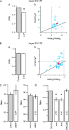
Loci of expression and receptor dependence of STDP in L2/3 P–P connections. (A) LTP is expressed presynaptically as demonstrated by (a) a significant decrease in PPR after induction of potentiation (b) CV analysis (n = 26). (B) Meanwhile, LTD is expressed postsynaptically as indicated by (a) the unchanged PPR after depression induction and (b) CV analysis (n = 26). (C) Both LTP and LTD are unaffected by application of CB1 receptor antagonist (2 μM AM251, n = 4 for LTP and n = 7 for LTD). (D) LTP requires NMDAR activation, whereas LTD is mGluR dependent: 1) LTD (n = 7) was not blocked in the presence of 50 μM APV, whereas LTP protocol (n = 4) induced LTD in the presence of 50 μM APV; 2) mGluR antagonists prevent LTD induction (n = 4). In (D), EPSPs were normalized to the mean baseline EPSP amplitude. In all experiments, train-protocols were used for plasticity induction.

Similar articles

Cited by

References

    1. Aizenman CD, Manis PB, Linden DJ. Polarity of long-term synaptic gain change is related to postsynaptic spike firing at a cerebellar inhibitory synapse. Neuron. 1998;21:827–835. - PubMed
    1. Bender VA, Bender KJ, Brasier DJ, Feldman DE. Two coincidence detectors for spike timing-dependent plasticity in somatosensory cortex. J Neurosci. 2006;26:4166–4177. - PMC - PubMed
    1. Bi G-Q, Poo M-M. Synaptic modifications in cultured hippocampal neurons: dependence on spike timing, synaptic strength, and postsynaptic cell type. J Neurosci. 1998;18:10464–10472. - PMC - PubMed
    1. Bi G-Q, Poo M-M. Synaptic modification by correlated activity: Hebb's postulate revisited. Annu Rev Neurosci. 2001;24:139–166. - PubMed
    1. Brasier DJ, Feldman DE. Synapse-specific expression of functional presynaptic NMDA receptors in rat somatosensory cortex. J Neurosci. 2008;28:2199–2211. - PMC - PubMed

Publication types