

MR imaging findings in autosomal recessive hereditary spastic paraplegia
- PMID: 19193756
- PMCID: PMC7051668
- DOI: 10.3174/ajnr.A1483
MR imaging findings in autosomal recessive hereditary spastic paraplegia
Abstract
Background and purpose: Hereditary spastic paraplegia (HSP) is a disorder characterized by degeneration of the corticospinal tracts and posterior column of the spinal cord. Previously described radiologic findings included nonspecific brain abnormalities such as brain atrophy and white matter lesions, as well as atrophy of the spinal cord. In our study, we aimed to better characterize brain and spine MR imaging findings in a series of patients with HSP.
Materials and methods: Nine patients from 4 different Lebanese families with the autosomal recessive form of HSP were included in the study. All patients underwent brain and whole-spine MR imaging. We assessed the presence of white matter abnormalities mainly along the corticospinal tracts, brain atrophy, thinning of the corpus callosum, and the presence of spinal cord atrophy or abnormal signal intensity.
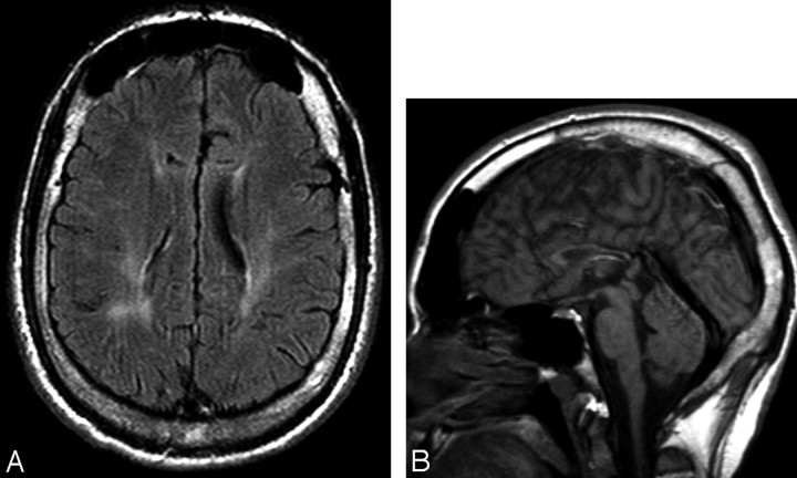
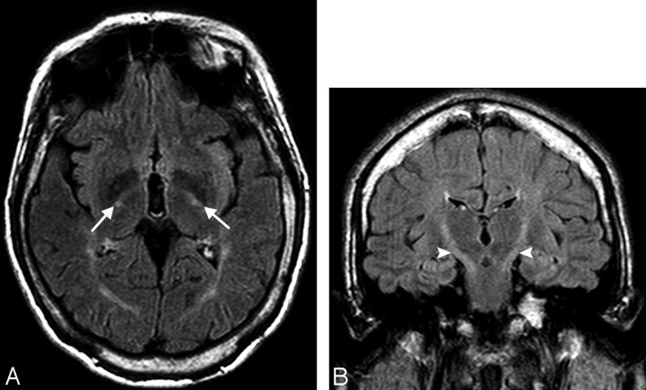
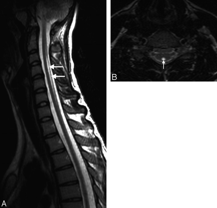
Results: Imaging revealed mild brain atrophy (44%), atrophy of the corpus callosum (55%), white matter lesions (67%), abnormal T2 high signal intensity in the posterior limb of the internal capsule (55%), and mild spinal cord atrophy (33%).
Conclusions: The MR imaging findings of HSP are nonspecific and variable; however, the most prominent features include atrophy of the corpus callosum, T2 signal intensity in the posterior limb of the internal capsule, and spinal cord atrophy.
Figures
Similar articles
-
Multimodal MRI-based study in patients with SPG4 mutations.PLoS One. 2015 Feb 6;10(2):e0117666. doi: 10.1371/journal.pone.0117666. eCollection 2015. PLoS One. 2015. PMID: 25658484 Free PMC article.
-
Spinal cord magnetic resonance imaging in autosomal dominant hereditary spastic paraplegia.Neuroradiology. 2005 Oct;47(10):730-4. doi: 10.1007/s00234-005-1415-3. Epub 2005 Sep 6. Neuroradiology. 2005. PMID: 16143870
-
Spinal Cord Gray and White Matter Damage in Different Hereditary Spastic Paraplegia Subtypes.AJNR Am J Neuroradiol. 2021 Mar;42(3):610-615. doi: 10.3174/ajnr.A7017. Epub 2021 Jan 21. AJNR Am J Neuroradiol. 2021. PMID: 33478946 Free PMC article.
-
Cerebrotendinous xanthomatosis (van Bogaert-Scherer-Epstein disease): CT and MR findings.AJNR Am J Neuroradiol. 1994 Oct;15(9):1721-6. AJNR Am J Neuroradiol. 1994. PMID: 7847220 Free PMC article. Review.
-
[Three patients of complicated form of autosomal recessive hereditary spastic paraplegia associated with hypoplasia of the corpus callosum].No To Shinkei. 1994 Oct;46(10):941-7. No To Shinkei. 1994. PMID: 7826709 Review. Japanese.
Cited by
-
Multiparametric 3T MRI evaluation of hereditary spastic paraplegia: A case report.Indian J Radiol Imaging. 2016 Jul-Sep;26(3):328-331. doi: 10.4103/0971-3026.190413. Indian J Radiol Imaging. 2016. PMID: 27857457 Free PMC article.
-
Selective Dorsal Rhizotomy for Treatment of Hereditary Spastic Paraplegia-Associated Spasticity in 37 Patients.Cureus. 2021 Sep 3;13(9):e17690. doi: 10.7759/cureus.17690. eCollection 2021 Sep. Cureus. 2021. PMID: 34650864 Free PMC article.
-
Evidence for autosomal recessive inheritance in SPG3A caused by homozygosity for a novel ATL1 missense mutation.Eur J Hum Genet. 2014 Oct;22(10):1180-4. doi: 10.1038/ejhg.2014.5. Epub 2014 Jan 29. Eur J Hum Genet. 2014. PMID: 24473461 Free PMC article.
-
Clinico-Investigative Profile of Hereditary Spastic Paraplegia in Children.Ann Indian Acad Neurol. 2019 Jul-Sep;22(3):341-344. doi: 10.4103/aian.AIAN_527_18. Ann Indian Acad Neurol. 2019. PMID: 31359954 Free PMC article.
-
Neuroimaging in Hereditary Spastic Paraplegias: Current Use and Future Perspectives.Front Neurol. 2019 Jan 16;9:1117. doi: 10.3389/fneur.2018.01117. eCollection 2018. Front Neurol. 2019. PMID: 30713518 Free PMC article. Review.
References
-
- Goldblatt J, Ballo R, Sachs B, et al. X-linked spastic paraplegia: evidence for homogeneity with a variable phenotype. Clin Genet 1989;35:116–20 - PubMed
-
- Durr A, Brice A, Serdaru M, et al. The phenotype of “pure” autosomal dominant spastic paraplegia. Neurology 1994;44:1274–77 - PubMed
-
- Hedera P, Eldevik OP, Maly P, et al. Spinal cord magnetic resonance imaging in autosomal dominant hereditary spastic paraplegia. Neuroradiology 2005;47:730–34 - PubMed
-
- Lesca G, Eymard-Pierre E, Santorelli FM, et al. Infantile ascending hereditary spastic paralysis (IAHSP): clinical features in 11 families. Neurology 2003;60:674–82 - PubMed
Publication types
MeSH terms
LinkOut - more resources
Full Text Sources
Medical