. 2009 Apr;83(8):3420-8.
doi: 10.1128/JVI.02623-08. Epub 2009 Feb 4.

MAVS self-association mediates antiviral innate immune signaling

Affiliations

MAVS self-association mediates antiviral innate immune signaling

Eric D Tang et al. J Virol. 2009 Apr.

Abstract

The innate immune system recognizes nucleic acids during viral infection and stimulates cellular antiviral responses. Intracellular detection of RNA virus infection is mediated by the RNA helicases RIG-I (retinoic acid inducible gene I) and MDA-5, which recognize viral RNA and signal through the adaptor molecule MAVS (mitochondrial antiviral signaling) to stimulate the phosphorylation and activation of the transcription factors IRF3 (interferon regulatory factor 3) and IRF7. Once activated, IRF3 and IRF7 turn on the expression of type I interferons, such as beta interferon. Interestingly, unlike other signaling molecules identified in this pathway, MAVS contains a C-terminal transmembrane (TM) domain that is essential for both type I interferon induction and localization of MAVS to the mitochondrial outer membrane. However, the role the MAVS TM domain plays in signaling remains unclear. Here we report the identification of a function for the TM domain in mediating MAVS self-association. The activation of RIG-I/MDA-5 leads to the TM-dependent dimerization of the MAVS N-terminal caspase recruitment domain, thereby providing an interface for direct binding to and activation of the downstream effector TRAF3 (tumor necrosis factor receptor-associated factor 3). Our results reveal a role for MAVS self-association in antiviral innate immunity signaling and provide a molecular mechanism for downstream signal transduction.


Figures
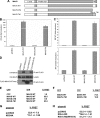
FIG. 1.
FIG. 1.
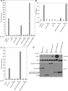
The TM domain is required for MAVS self-association. (A) Schematic representation of MAVS mutant constructs. WT, wild type. (B) Luciferase reporter assay for HEK293T cells cotransfected with wild-type or mutant MAVS and an IFN-β promoter reporter construct, pLUC-IFN-β. (C) Luciferase reporter assay for HEK293T cells cotransfected with wild-type or mutant MAVS and the pLUC-PRD(III-I)3 or pLUC-PRD(II)2 reporter construct. (D) Coimmunoprecipitation assay with lysates prepared from HEK293T cells cotransfected with FLAG-tagged wild-type or mutant (mut) MAVS and HA-tagged wild-type MAVS. Anti-FLAG immunoprecipitates (IP) or lysates were immunoblotted for HA- or FLAG-tagged MAVS constructs. (E, F) FRET analysis of MAVS−/− MEFs cotransfected with YFP, YFP-tagged wild-type MAVS, or YFP-MAVSΔTM and either CFP, CFP-tagged MAVS, or CFP-MAVSΔTM constructs. The percentage of cells that coexpressed both CFP and YFP and displayed FRET was measured by flow cytometry. (G, H) FRET analysis of MAVS−/− MEFs transfected with CFP-MAVS and YFP-MAVS along with NS3/4A, RIG-IΔRD, or RIG-IΔRD(W167A). The percentage of cells that expressed both CFP and YFP and displayed FRET was measured (mean ± standard deviation). Samples were analyzed in triplicate. *, P values of <0.01 for comparison with pcDNA3 (n = 2); **, P values of <0.0001 for comparison with pcDNA3 (n = 3).
FIG. 2.
FIG. 2.
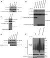
Restoration of MAVS signaling in the absence of a TM domain by induced self-association. (A) Luciferase reporter assay for HEK293T cells transfected with wild-type (WT) or mutant MAVS and an IFN-β reporter, pLUC-IFN-β. AP1510 was added 12 h prior to cell lysis. (B) Human IFN-β enzyme-linked immunosorbent assays at 36 h with tissue culture supernatants of HEK293 cells transfected with wild-type MAVS or mutant MAVS constructs. Samples were prepared in duplicate. The concentration of IFN-β is shown (mean ± standard deviation). AP1510 was added 12 h prior to analysis. ND, not detectable. (C) Luciferase reporter assay for HEK293T cells transfected with wild-type or mutant MAVS and pLuc-PRD(III-I)3. AP1510 was added 12 h prior to cell lysis. (D) Immunoblotting for phosphorylated IRF3 (Ser 396) from anti-HA immunoprecipitates (IP) prepared from lysates of HEK293T cells transfected with FLAG-tagged MAVS, FPK3, or MAVS-FPK3 fusions along with HA-IRF3. Lysates were immunoblotted for HA-IRF3 and FLAG-tagged proteins. (E) Immunoblotting for phosphorylated IRF3 (Ser 396) from anti-HA immunoprecipitates in lysates prepared from HEK293T cells transfected with FLAG-tagged MAVS CARD-FPK3. AP1510 was added prior to lysis for various times. Images representing both short and long exposures are shown. Anti-HA immunoprecipitates were also immunoblotted for HA-IRF3. Lysates were immunoblotted for MAVS CARD-FPK3. (F) Monitoring for VSV-GFP replication by flow cytometry with infected HEK293 cells transiently transfected with MAVS constructs. The percentage of cells positive for GFP is indicated (mean ± standard deviation). Virus infection was performed 48 h following transfection. AP1510 was added 12 h prior to infection. Regular culture medium was added after infection. *, P values of <0.001 for comparison with pcDNA3 (n = 3); **, P values of <0.00001 for comparison with pcDNA3 (n = 3). (G) Luciferase reporter assay for HEK293T cells cotransfected with FLAG-tagged wild-type MAVS, MAVSΔTM, or MAVSΔTMx2 and an IRF3/7 reporter, pLuc-PRD(III-I)3. Lysates were immunoblotted for FLAG-tagged proteins. (H) Immunoblotting for phosphorylated IRF3 (Ser 396) and phosphorylated IκBα (Ser 32) from anti-HA immunoprecipitates in lysates prepared from HEK293T cells transfected with FLAG-tagged wild-type MAVS, MAVSΔTM, or MAVSΔTMx2 along with HA-IRF3. Immunoprecipitates were also immunoblotted for HA-IRF3. (I) Cross-linking of presumptive MAVS homodimer from purified mitochondria isolated from HEK293T cells transfected with poly(I:C). Mitochondria were incubated with vehicle (dimethyl sulfoxide) or BMH, and total proteins were separated by SDS-PAGE and probed for endogenous MAVS. The arrowhead indicates 150-kDa presumptive MAVS homodimeric species. Asterisks indicate nonspecific reactive bands. The bottom panel shows an image representing a short exposure, demonstrating monomeric, 75-kDa full-length MAVS.
FIG. 2.
FIG. 2.
Restoration of MAVS signaling in the absence of a TM domain by induced self-association. (A) Luciferase reporter assay for HEK293T cells transfected with wild-type (WT) or mutant MAVS and an IFN-β reporter, pLUC-IFN-β. AP1510 was added 12 h prior to cell lysis. (B) Human IFN-β enzyme-linked immunosorbent assays at 36 h with tissue culture supernatants of HEK293 cells transfected with wild-type MAVS or mutant MAVS constructs. Samples were prepared in duplicate. The concentration of IFN-β is shown (mean ± standard deviation). AP1510 was added 12 h prior to analysis. ND, not detectable. (C) Luciferase reporter assay for HEK293T cells transfected with wild-type or mutant MAVS and pLuc-PRD(III-I)3. AP1510 was added 12 h prior to cell lysis. (D) Immunoblotting for phosphorylated IRF3 (Ser 396) from anti-HA immunoprecipitates (IP) prepared from lysates of HEK293T cells transfected with FLAG-tagged MAVS, FPK3, or MAVS-FPK3 fusions along with HA-IRF3. Lysates were immunoblotted for HA-IRF3 and FLAG-tagged proteins. (E) Immunoblotting for phosphorylated IRF3 (Ser 396) from anti-HA immunoprecipitates in lysates prepared from HEK293T cells transfected with FLAG-tagged MAVS CARD-FPK3. AP1510 was added prior to lysis for various times. Images representing both short and long exposures are shown. Anti-HA immunoprecipitates were also immunoblotted for HA-IRF3. Lysates were immunoblotted for MAVS CARD-FPK3. (F) Monitoring for VSV-GFP replication by flow cytometry with infected HEK293 cells transiently transfected with MAVS constructs. The percentage of cells positive for GFP is indicated (mean ± standard deviation). Virus infection was performed 48 h following transfection. AP1510 was added 12 h prior to infection. Regular culture medium was added after infection. *, P values of <0.001 for comparison with pcDNA3 (n = 3); **, P values of <0.00001 for comparison with pcDNA3 (n = 3). (G) Luciferase reporter assay for HEK293T cells cotransfected with FLAG-tagged wild-type MAVS, MAVSΔTM, or MAVSΔTMx2 and an IRF3/7 reporter, pLuc-PRD(III-I)3. Lysates were immunoblotted for FLAG-tagged proteins. (H) Immunoblotting for phosphorylated IRF3 (Ser 396) and phosphorylated IκBα (Ser 32) from anti-HA immunoprecipitates in lysates prepared from HEK293T cells transfected with FLAG-tagged wild-type MAVS, MAVSΔTM, or MAVSΔTMx2 along with HA-IRF3. Immunoprecipitates were also immunoblotted for HA-IRF3. (I) Cross-linking of presumptive MAVS homodimer from purified mitochondria isolated from HEK293T cells transfected with poly(I:C). Mitochondria were incubated with vehicle (dimethyl sulfoxide) or BMH, and total proteins were separated by SDS-PAGE and probed for endogenous MAVS. The arrowhead indicates 150-kDa presumptive MAVS homodimeric species. Asterisks indicate nonspecific reactive bands. The bottom panel shows an image representing a short exposure, demonstrating monomeric, 75-kDa full-length MAVS.
FIG. 3.
FIG. 3.
Self-association of MAVS allows for TRAF3 binding. (A) Coimmunoprecipitation assay with lysates prepared from HEK293T cells cotransfected with FLAG-tagged MAVS constructs together with HA-tagged TRAF3. Anti-FLAG-immunoprecipitates (IP) were immunoblotted for HA-TRAF3 or FLAG-MAVS proteins. Lysates were immunoblotted for HA-TRAF3. (B) Coimmunoprecipitation assay with lysates prepared from HEK293T cells cotransfected with FLAG-tagged MAVS constructs together with HA-tagged TRAF3. Anti-FLAG immunoprecipitates were immunoblotted for HA-TRAF3 or FLAG-MAVS proteins. Lysates were immunoblotted for HA-TRAF3. AP1510 was added 12 h prior to cell lysis. WT, wild type. (C) Coimmunoprecipitation assay with lysates prepared from HEK293T cells cotransfected with FLAG-MAVSΔTM or FLAG-MAVSΔTMx2 together with HA-tagged TRAF3. Anti-FLAG-immunoprecipitates were immunoblotted for HA-TRAF3 or FLAG-MAVS proteins. Lysates were immunoblotted for HA-TRAF3. (D) Coimmunoprecipitation assay with lysates prepared from HEK293T cells cotransfected with FLAG-tagged MAVS constructs together with HA-tagged TRAF3. Anti-FLAG immunoprecipitates were immunoblotted for HA-TRAF3 or FLAG-MAVS proteins. Lysates were immunoblotted for HA-TRAF3. mut, mutant. (E) Immunoblotting for Ub-conjugated TRAF3 from HEK293T cells cotransfected with FLAG-tagged FPK3, MAVS CARD-FPK3, or MAVS CARDmt-FPK3 along with AU1-Ub and HA-TRAF3. Anti-HA immunoprecipitates were immunoblotted for AU1-Ub conjugates or HA-TRAF3. Lysates were immunoblotted for FLAG-tagged proteins. AP1510 was added 12 h prior to cell lysis.

Similar articles

Cited by

References

    1. Akira, S., S. Uematsu, and O. Takeuchi. 2006. Pathogen recognition and innate immunity. Cell 124783-801. - PubMed
    1. Baril, M., M. E. Racine, F. Penin, and D. Lamarre. 2009. MAVS dimer is a crucial signaling component of innate immunity and the target of hepatitis C virus NS3/4A protease. J. Virol. 831299-1311. - PMC - PubMed
    1. Borden, E. C., G. C. Sen, G. Uze, R. H. Silverman, R. M. Ransohoff, G. R. Foster, and G. R. Stark. 2007. Interferons at age 50: past, current and future impact on biomedicine. Nat. Rev. Drug Discov. 6975-990. - PMC - PubMed
    1. Chin, C. N., J. N. Sachs, and D. M. Engelman. 2005. Transmembrane homodimerization of receptor-like protein tyrosine phosphatases. FEBS Lett. 5793855-3858. - PubMed
    1. Choi, S., E. Lee, S. Kwon, H. Park, J. Y. Yi, S. Kim, I. O. Han, Y. Yun, and E. S. Oh. 2005. Transmembrane domain-induced oligomerization is crucial for the functions of syndecan-2 and syndecan-4. J. Biol. Chem. 28042573-42579. - PubMed

Publication types