. 2009 Apr;83(8):3704-18.
doi: 10.1128/JVI.01384-08. Epub 2009 Feb 4.

O-linked N-acetylglucosaminylation of Sp1 inhibits the human immunodeficiency virus type 1 promoter

Affiliations

O-linked N-acetylglucosaminylation of Sp1 inhibits the human immunodeficiency virus type 1 promoter

Ramona Jochmann et al. J Virol. 2009 Apr.

Abstract

Human immunodeficiency virus type 1 (HIV-1) gene expression and replication are regulated by the promoter/enhancer located in the U3 region of the proviral 5' long terminal repeat (LTR). The binding of cellular transcription factors to specific regulatory sites in the 5' LTR is a key event in the replication cycle of HIV-1. Since transcriptional activity is regulated by the posttranslational modification of transcription factors with the monosaccharide O-linked N-acetyl-D-glucosamine (O-GlcNAc), we evaluated whether increased O-GlcNAcylation affects HIV-1 transcription. In the present study we demonstrate that treatment of HIV-1-infected lymphocytes with the O-GlcNAcylation-enhancing agent glucosamine (GlcN) repressed viral transcription in a dose-dependent manner. Overexpression of O-GlcNAc transferase (OGT), the sole known enzyme catalyzing the addition of O-GlcNAc to proteins, specifically inhibited the activity of the HIV-1 LTR promoter in different T-cell lines and in primary CD4(+) T lymphocytes. Inhibition of HIV-1 LTR activity in infected T cells was most efficient (>95%) when OGT was recombinantly overexpressed prior to infection. O-GlcNAcylation of the transcription factor Sp1 and the presence of Sp1-binding sites in the LTR were found to be crucial for this inhibitory effect. From this study, we conclude that O-GlcNAcylation of Sp1 inhibits the activity of the HIV-1 LTR promoter. Modulation of Sp1 O-GlcNAcylation may play a role in the regulation of HIV-1 latency and activation and links viral replication to the glucose metabolism of the host cell. Hence, the establishment of a metabolic treatment might supplement the repertoire of antiretroviral therapies against AIDS.


Figures
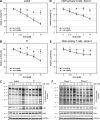
FIG. 1.
FIG. 1.
GlcN inhibits HIV-1 transcription in lymphocytes. Jurkat cells (A), T1 cells (B), and primary CD4+ T cells from two different donors (D and E) were infected with VSV-G env pseudotyped HIV-1NL4-3LucR-E- and cultured in the absence (0 mM) or presence of different concentrations of GlcN (0.25 mM, 1 mM, 4 mM, and 16 mM) for 24 h. Subsequently, HIV-1 LTR-driven luciferase activity was measured (solid lines). The effect of GlcN on cytotoxicity and proliferation was monitored by MTT assay (dashed lines). The results are presented in terms of percent activities of untreated control cells. The means ± standard deviations from triplicate determinations are indicated. P values are calculated in comparison with control: *, P ≤ 0.05; **, P ≤ 0.01. (C and F) Western blot analyses of O-GlcNAcylated proteins and OGT in Jurkat and T1 cells (C) and in primary CD4+ T lymphocytes (F). Actin and GAPDH served as loading controls.
FIG. 2.
FIG. 2.
GlcN inhibits HIV-1 LTR promoter activity and increases O-GlcNAcylation in HeLa cells. (A) HeLa-Tat-III/LTR/d1EGFP cells were either left untreated (0 mM) or stimulated for 5 h with increasing concentrations of GlcN (0.25 mM, 1 mM, 4 mM, and 16 mM). The HIV-1 LTR activity was assessed by measuring the d1EGFP fluorescence intensity (FI) with flow cytometry analyses. The shift of the fluorescence emission peak (inset) and the bar diagram of the respective geometric mean values compared to those for unstimulated control cells are shown. The means ± standard deviations calculated from triplicate determinations are indicated. P values are given for comparison with control: n.s., not significant; **, P ≤ 0.01; ***, P ≤ 0.001. (B) Nuclear and cytosolic fractions of unstimulated (control) or stimulated (16 mM GlcN) HeLa-Tat-III/LTR/d1EGFP cells were analyzed by Western blotting to detect O-GlcNAcylated proteins. Arrowheads mark proteins whose O-GlcNAcylation patterns are increased upon GlcN treatment. Staining of OGT (an example of an O-GlcNAcylated protein) and histone H1 (non-O-GlcNAcylated) shows that protein expression levels were not altered. Staining of GAPDH and lamin A/C demonstrates successful fractionation.
FIG. 3.
FIG. 3.
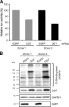
OGT inhibits HIV-1 LTR promoter activity in infected primary CD4+ T cells. Primary CD4+ T cells from two different donors were infected with VSV-G env pseudotyped HIV-1NL4-3LucR-E-. At 36 h postinfection, cells were electroporated with in vitro-transcribed polyadenylated mRNA encoding either EGFP or OGT. (A) At 8 h postelectroporation, luciferase activity was measured. (B) The O-GlcNAcylation pattern and overexpression of EGFP and OGT were verified by Western blotting. Staining of GAPDH demonstrates equal loading of proteins.
FIG. 4.
FIG. 4.
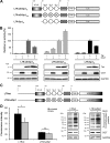
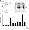
The presence of Sp1-binding sites in the HIV-1 LTR promoter is required for OGT-mediated inhibition. (A) Schematic representation of the promoter constructs used in the luciferase assay in panel B. The truncated promoters lack the upstream modulatory region but contain the NF-κB- and/or Sp1-binding sites (either combined or isolated), as well as a TATA box and the TAR element for Tat-mediated activation. Quartered circles represent mutated Sp1-binding sites. Numbers reflect the positions on the wild-type LTRLAI. (B) Promoter activities were detected by measuring the luciferase activity in the cell lysates of HEK 293T cells transiently transfected with the reporter constructs (LTR-κB-Sp1wt, LTR-κB-Sp1mut, and LTR-Sp1wt) together with plasmids coding for OGT and Tat as indicated. The total DNA amount was adjusted with pcDNA4. The values were normalized to the total amount of protein and are presented in terms of percentages of the corresponding Tat-induced promoter activity. The means ± standard deviations from triplicate determinations are shown (upper panel). Western blot analyses of Tat and OGT in transfected cells are shown in the lower panel. GAPDH staining demonstrates that equal amounts of protein were loaded. (C) Schematic representation of the full-length wild-type and full-length Sp1-mutated promoter constructs used for the stable transfection of HEK 293T cells. (D) HEK 293T cells were stably transfected with d2EGFP either under the control of the wild-type HIV-1 LTR (LTRwt) or under the control of the LTR containing mutated Sp1-binding sites in the basal promoter (LTRmutSp1). The effect of GlcN (16 mM) on the HIV-1 LTR was analyzed by flow cytometry, and the mean fluorescence intensity is represented in the bar diagram. The means ± standard deviations were calculated from duplicate determinations of three independent clones with similar fluorescence intensities. P values are given for comparison with control: *, P ≤ 0.05; n.s., not significant. Western blot analyses of one representative clone verify the increased O-GlcNAcylation pattern upon GlcN treatment and constant OGT expression. GAPDH staining demonstrates that equal amounts of protein were loaded.
FIG. 5.
FIG. 5.
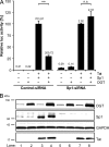
Sp1 is crucial for the inhibitory effect of OGT on the HIV-1 LTR. (A) HIV-1 LTR activity was measured by luciferase assay after cotransfection of HEK 293T cells with an LTR-Sp1wt reporter construct along with Tat-and Sp1-encoding plasmids in the absence or presence of an OGT-encoding vector together with 10 nM control siRNA or an siRNA specifically targeting Sp1. Total DNA amount was adjusted with pcDNA4. The relative light units normalized to the total protein content indicate the means ± standard deviations from triplicate determinations (values above the bars). The results were normalized for each siRNA data set individually and are presented in terms of percentages of control (Tat- and Sp1-induced LTR-Sp1wt activity). P values are given for comparison with control: **, P ≤ 0.01; n.s., not significant. (B) Western blot analyses of the lysates verify the knockdown of Sp1 by siRNA and prove that the expression of OGT and Tat remains unaffected upon silencing of Sp1. The GAPDH staining demonstrates that equal amounts of protein were loaded.
FIG. 6.
FIG. 6.
O-GlcNAcylation of Sp1 selectively inhibits HIV-1 LTR promoter activity in a dose-dependent manner. (A) HEK 293T cells were transfected with control vector (CV), Sp1, or Sp1- and OGT-expressing vectors. Total DNA amount was adjusted with pcDNA4. Expression of transfected plasmids and O-GlcNAcylation were assessed via Western blotting of cell lysates (input). Immunoprecipitation of Sp1 (Sp1-IP) and O-GlcNAcylation of Sp1 were verified by Western blotting of Sp1-IPs with anti-Sp1 and anti-O-GlcNAc antibodies, respectively. (B) The effects of OGT on the LTR-Sp1wt and the control promoter EF1α in HEK 293T cells cotransfected with the reporter and an Sp1-encoding plasmid were analyzed by luciferase assay. (C) Luciferase activity of HEK 293T cells transiently cotransfected with the LTR-Sp1wt reporter construct along with Tat- and Sp1-encoding plasmids and increasing concentrations of an OGT-encoding vector (0.05 μg, 0.2 μg, and 1 μg). The total DNA amount in panels B and C was adjusted with pcDNA4. The values were normalized to the total amount of protein and are presented in terms of percentages of control (Sp1 induced [B] and Tat- and Sp1-induced [C] promoter activity). The means ± standard deviations from triplicate determinations are indicated. P values are given for comparison with control: n.s., not significant; *, P ≤ 0.05; **, P ≤ 0.01.
FIG. 7.
FIG. 7.
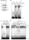
OGT does not interfere with Sp1 expression and DNA binding of Sp1. (A) HEK 293T cells were transfected with an Sp1-encoding plasmid, along with an OGT-encoding plasmid or control plasmid. Protein expression was detected in nuclear protein extracts via Western blotting. Staining of lamin A/C was used as a loading control. (B) Electrophoretic mobility shift assays were carried out with 32P-end-labeled double-stranded oligonucleotides corresponding to the Sp1-binding sites in the HIV-1 LTR promoter (wt LTR-Sp1) or with oligonucleotides containing mutated Sp1-binding sites in order to prevent binding (mut LTR-Sp1). One representative gel shift assay out of three is shown. Reactions were performed either without nuclear extracts (free) or with lysates from cells transfected with an Sp1-encoding plasmid alone (Sp1) or in combination with an OGT-encoding plasmid (Sp1+OGT). The Sp1-oligonucleotide complex is indicated as Sp1-C. (C) Competition experiments were carried out with 10-, 20-, and 50-fold molar excesses of unlabeled wild-type (wt LTR-Sp1) or mutated (mut LTR-Sp1) oligonucleotides. (D) Supershift analyses were performed using 1 μg, 2 μg, or 5 μg anti-Sp1, anti-O-GlcNAc, or anti-OGT antibodies as well as 5 μg anti-IgG or anti-IgM as an isotype control.
FIG. 8.
FIG. 8.
Sp1 O-GlcNAcylation is necessary for the inhibition of the HIV-1 LTR. (A) Generation of a plasmid encoding an OGT rescue mutant in order to escape silencing by the siRNA targeting wild-type OGT: six silent mutations (bold) were introduced into the siRNA-binding sequence of OGT. (B) Western blot analyses of wild-type (wt) and rescue mutant (res) OGT were performed after transfection with the siRNA specifically targeting wtOGT. Detection of O-GlcNAcylation levels was used as a control for the functionality of the rescue mutant. Immunodetection of GAPDH demonstrates that equal amounts of protein were loaded. (C) HEK 293T cells were transfected with the reporter construct LTR-Sp1wt along with plasmids encoding Sp1 and OGT (wild type or rescue mutant). Total DNA amount was adjusted with pcDNA4. Control siRNA or siRNA targeting wtOGT (10 nM) was cotransfected, and the luciferase activity was measured. The values were adjusted to the total amount of protein and indicate the means ± standard deviations from triplicate determinations. The results are presented in terms of percentages of control (Sp1-induced LTR activity).
FIG. 9.
FIG. 9.
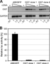
OGT overexpression prior to infection amplifies the inhibitory effect on HIV-1 replication. (A) Jurkat cells were stably transfected with plasmids encoding either d2EGFP or OGT. Expression of recombinantly expressed OGT mRNA was confirmed by RT-PCR (upper panel) using specific primers. Amplification of GAPDH served as a loading control (lower panel). Cellular cDNA was subjected to the amplification reactions in increasing dilutions: undiluted (1), 1:5, and 1:10. Control reactions were carried out in the absence of reverse transcriptase (−RT) with undiluted cDNA template. (B) Jurkat cells stably expressing d2EGFP or OGT were infected with VSV-G env pseudotyped HIV-1NL4-3LucR-E-. Luciferase activity was measured 48 h after infection.

Similar articles

Cited by

References

    1. Adachi, A., H. E. Gendelman, S. Koenig, T. Folks, R. Willey, A. Rabson, and M. A. Martin. 1986. Production of acquired immunodeficiency syndrome-associated retrovirus in human and nonhuman cells transfected with an infectious molecular clone. J. Virol. 59284-291. - PMC - PubMed
    1. Berkhout, B., and K. T. Jeang. 1992. Functional roles for the TATA promoter and enhancers in basal and Tat-induced expression of the human immunodeficiency virus type 1 long terminal repeat. J. Virol. 66139-149. - PMC - PubMed
    1. Blough, H. A., and R. L. Giuntoli. 1979. Successful treatment of human genital herpes infections with 2-deoxy-D-glucose. JAMA 2412798-2801. - PubMed
    1. Bonehill, A., C. Heirman, S. Tuyaerts, A. Michiels, K. Breckpot, F. Brasseur, Y. Zhang, P. Van Der Bruggen, and K. Thielemans. 2004. Messenger RNA-electroporated dendritic cells presenting MAGE-A3 simultaneously in HLA class I and class II molecules. J. Immunol. 1726649-6657. - PubMed
    1. Bonello, M. R., and L. M. Khachigian. 2004. Fibroblast growth factor-2 represses platelet-derived growth factor receptor-alpha (PDGFR-alpha) transcription via ERK1/2-dependent Sp1 phosphorylation and an atypical cis-acting element in the proximal PDGFR-alpha promoter. J. Biol. Chem. 2792377-2382. - PubMed

Publication types

Substances