

doi: 10.1073/pnas.0810682106.
Epub 2009 Feb 4.
Efficient gene therapy-based method for the delivery of therapeutics to primate cortex
Affiliations
- PMID: 19193857
- PMCID: PMC2650169
- DOI: 10.1073/pnas.0810682106
Item in Clipboard
Efficient gene therapy-based method for the delivery of therapeutics to primate cortex
Proc Natl Acad Sci U S A.
.
Abstract
Transduction of the primate cortex with adeno-associated virus (AAV)-based gene therapy vectors has been challenging, because of the large size of the cortex. We report that a single infusion of AAV2 vector into thalamus results in widespread expression of transgene in the cortex through transduction of widely dispersed thalamocortical projections. This finding has important implications for the treatment of certain genetic and neurodegenerative diseases.
Conflict of interest statement
The authors declare no conflict of interest.
Figures
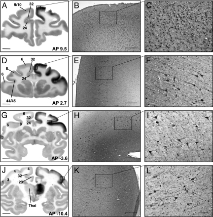
Distribution of GDNF protein after AAV2-GDNF infusion into the thalamus. (A) GDNF expression detected by immunohistochemistry staining in the prefrontal cortex ipsilateral to thalamic infusion. (B and C) Large numbers of nonpyramidal GDNF-positive neurons can be seen across multiple layers in the prefrontal cortex (areas 9 and 10). (D–G) GDNF expression in the premotor cortex (area 6), frontal eye fields (area 8), Broca's area (areas 44 and 45), and cingulate cortex (areas 23, 24, and 32). Pyramidal neurons in laminae V and VI of the premotor cortex (E and F) and frontal eye fields (H and I) expressing GDNF. Strong GDNF-immunopositive staining is evident in the cortical layers above the pyramidal neurons. (J) Intensive GDNF-positive staining in the infused thalamus and cortical GDNF expression in the somatosensory cortex (areas 1, 2, and 3) and primary motor cortex (area 4). (K and L) GDNF-positive neurons in laminae V and VI of the somatosensory cortex. AP, anterior–posterior distance (in mm) from the bregma. Thal, thalamus. Numbers indicate different cortical areas referenced in the text. (Scale bars: 5 mm for A, D, G, and J; 500 μm for B, E, H, and K; 100 μm for C, F, I, and L.)
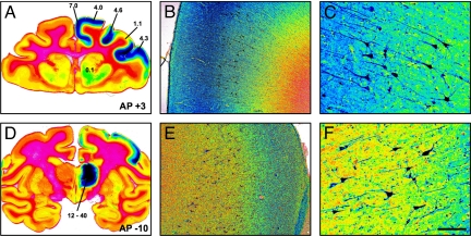
Level of GDNF protein expression after infusion of AAV2-GDNF into the right thalamus. Pseudocolor images of GDNF immunohistochemistry-stained sections showing the gradients of GDNF distribution in both the thalamus and cortex. Blue represents the highest intensity of DAB staining; red, the lowest intensity. The numbers in panels A and D represent the level of GDNF protein (μg of GDNF per mg of total protein) in different areas of the brain measured by ELISA from an adjacent tissue block. In panels B, C, E, and F, higher magnification of the cortex shows the high intensity of GDNF staining in lamina III/IV and the high cytoplasmic presence of GDNF in lamina V/VI pyramidal neurons. AP, anterior–posterior distance (in mm) from the bregma. (Scale bars: 10 mm in A and D; 500 μm in B and E; 100 μm in C and F.)
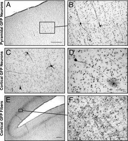
Cortical expression of GFP after infusion of AAV2-GFP to the left thalamus. Individual GFP-immunopositive neurons are seen within different areas of the cortex. (A and B) Cortical pyramidal neurons are the predominant type of GFP-positive neuron. (C and D) Neurons without pyramidal morphology also are seen in the cortex, including basket-like neurons (C) and glia-like cells (D). (E and F) Extensive GFP-positive fiber networks also are seen in the frontal cortex. (Scale bars: 500 μm in A and E; 100 μm in B and C; 50 μm in D and F.)
References
-
- Vite CH, et al. Effective gene therapy for an inherited CNS disease in a large animal model. Ann Neurol. 2005;57:355–364. - PubMed
-
- Vite CH, Passini MA, Haskins ME, Wolfe JH. Adeno-associated virus vector-mediated transduction in the cat brain. Gene Ther. 2003;10:1874–1881. - PubMed
-
- Kaplitt MG, et al. Safety and tolerability of gene therapy with an adeno-associated virus (AAV) borne GAD gene for Parkinson's disease: An open-label, phase I trial. Lancet. 2007;369:2097–2105. - PubMed
-
- Fiandaca M, Forsayeth J, Bankiewicz K. Current status of gene therapy trials for Parkinson's disease. Exp Neurol. 2008;209:51–57. - PubMed
-
- Eberling JL, et al. Results from a phase I safety trial of hAADC gene therapy for Parkinson disease. Neurology. 2008;70:1980–1983. - PubMed
Publication types
MeSH terms
Substances
Grants and funding
LinkOut - more resources
Full Text Sources
Other Literature Sources
Medical
