. 2009 Apr;296(4):C724-34.
doi: 10.1152/ajpcell.00409.2008. Epub 2009 Feb 4.

Attenuation of proliferation and migration of retinal pericytes in the absence of thrombospondin-1

Affiliations

Attenuation of proliferation and migration of retinal pericytes in the absence of thrombospondin-1

Elizabeth A Scheef et al. Am J Physiol Cell Physiol. 2009 Apr.

Abstract

Perivascular supporting cells, including vascular smooth muscle cells (VSMCs) and pericytes (PCs), provide instructive signals to adjacent endothelial cells helping to maintain vascular homeostasis. These signals are provided through direct contact and by the release of soluble factors by these cells. Thrombospondin (TSP)1 is a matricellular protein and an autocrine factor for VSMCs. TSP1 activity, along with that of PDGF, regulates VSMC proliferation and migration. However, the manner in which TSP1 and PDGF impact retinal PC function requires further investigation. In the present study, we describe, for the first time, the isolation and culture of retinal PCs from wild-type (TSP1(+/+)) and TSP1-deficient (TSP1(-/-)) immortomice. We showed that these cells express early and mature markers of PCs, including NG2, PDGF receptor-beta, and smooth muscle actin as well as desmin, calbindin, and mesenchymal stem cell markers. These cells were successfully passaged and maintained in culture for several months without significant loss of expression of these markers. TSP1(+/+) PCs proliferated at a faster rate compared with TSP1(-/-) PCs. In addition, TSP1(+/+) PCs, like VSMCs, responded to PDGF-BB with enhanced migration and proliferation. In contrast, TSP1(-/-) PCs failed to respond to the promigratory and proliferative activity of PDGF-BB. This may be attributed, at least in part, to the limited interaction of PDGF-BB with TSP1 in null cells, which is essential for PDGF proliferative and migratory action. We observed no significant differences in the rates of apoptosis in these cells. TSP1(-/-) PCs were also less adherent, expressed increased levels of TSP2 and fibronectin, and had decreased amounts of N-cadherin and alpha(v)beta(3)-integrin on their surface. Thus, TSP1 plays a significant role in retinal PC proliferation and migration impacting retinal vascular development and homeostasis.


Figures

Fig. 1.
Fig. 1.
Isolation and characterization of retinal pericytes (PCs). Top left: morphology of wild-type [thrombosporin (TSP)1+/+] and TSP1-deficient (TSP1−/−) mouse retinal PCs cultured on uncoated plates. Magnification: ×40. Bottom: expression of PC markers in TSP1+/+ (left) and TSP1−/− (right) PCs determined by FACS analysis in early passage [passage 5 (P5)] or later passage [passage 22 (P22)]. The shaded traces show staining in the absence of primary antibody. Please note the decreased levels of N-cadherin (N-Cad) in TSP1−/− PCs. Expression of NG2 (top right) varied between isolations and was affected by the confluency of cultures. PDGFRβ, PDGF receptor-β; SMA, smooth muscle actin. These experiments were repeated with three isolations of PCs with similar results.
Fig. 2.
Fig. 2.
Retinal PCs expressed mature PC markers. TSP1+/+ and TSP1−/− retinal PCs were cultured on glass slides and stained with specific antibodies to desmin and calbindin as described in materials and methods. Please note that the expression of these markers was minimally affected by the lack of TSP1. These experiments were repeated with three isolations of PCs with similar results.
Fig. 3.
Fig. 3.
Retinal PCs expressed other vascular cell markers. Expression levels of various markers were determined by FACS analysis using specific antibodies as described in materials and methods. The shaded traces show staining in the absence of primary antibody. Please note the lack of PECAM-1 expression, an endothelial cell marker, in retinal PCs, as expected. TSP1+/+ PCs expressed lower levels of endoglin. Expression levles of CD36, VCAM-1, and ICAM-1 were similar in TSP1+/+ and TSP1−/− retinal PCs. These experiments were repeated with three isolations of PCs with similar results.
Fig. 4.
Fig. 4.
Retinal PCs expressed mesenchymal stem cell markers. Expression levels of various markers were determined by FACS analysis using specific antibodies as described in materials and methods. The shaded traces show staining in the absence of primary antibody. Please note the expression of Sca1, CD11b, CD45, and CD90 in retinal PCs. Expression levels of CD11b, CD45, and CD90 were lower in TSP1−/− compared with TSP1+/+ PCs. These cells did not express CD73. These experiments were repeated with three isolations of PCs with similar results.
Fig. 5.
Fig. 5.
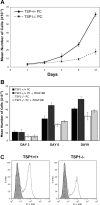
TSP1−/− PCs proliferated at a slower rate. The rate of cell proliferation was determined by counting the number of cells for 10 days under regular growth conditions (A) or in the presence of PDGF-BB (B) as described in materials and methods. Please note the significant decrease in the rate of proliferation of TSP1−/− PCs (n = 3, *P < 0.05). TSP1−/− PCs also failed to respond to the proliferative effect of PDGF-BB (n = 3, P > 0.05), whereas TSP1+/+ PC exhibited a significant increase in the rate of proliferation in response to PDGF-BB (n = 3, *P < 0.05). C: rates of cell proliferation in TSP1+/+ and TSP1−/− PCs were also evaluated by 5-ethynyl-2′-deoxyuridine (EdU) incorporation as described in materials and methods. A significant decrease in the percentage of PCs that incorporated EdU was observed in the absence of TSP1 (see text for quantifications). TSP1−/− PCs also failed to respond to the stimulatory effects of PDGF-BB in EdU labeling compared with TSP1+/+ PC (not shown).
Fig. 6.
Fig. 6.
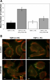
TSP1−/− retinal PCs did not respond to the promigratory effect of PDGF-BB. Migration of TSP1+/+ and TSP1−/− retinal PCs was determined in transwell assays with and without PDGF-BB as described in materials and methods. No significant differences in the basal migration of TSP1−/− PCs was observed compared with TSP1+/+ PCs. A significant increase in the migration of TSP1+/+ PCs in response to PDGF-BB was observed (n = 3, *P < 0.05). TSP1−/− retinal PCs did not show a significant response to the promigratory effects of PDGF-BB (n = 3, P > 0.05). The migratory characteristic of TSP1+/+ PCs in response to PDGF-BB was also demonstrated by staining with phalloidin (actin) and vinculin (focal adhesions). Please note the morphology changes in TSP1+/+ PCs incubated with PDGF-BB compared with untreated or TSP1−/− PCs with and without PDGF-BB. TSP1+/+ PCs incubated with PDGF-BB showed elongated morphology with numerous peripheral focal adhesions localizing to lamellipodia extensions along with increased actin stress fibers.
Fig. 7.
Fig. 7.
TSP1−/− retinal PCs were less adherent on fibronectin and vitronectin. The adhesion characteristics of retinal PCs to various extracellular matrix (ECM) proteins were determined as described in materials and methods. Please note the decreased adhesion to fibronectin and vitronectin in TSP1−/− PCs compared with TSP1+/+ PC. Retinal PCs adhered poorly to collagen and laminin. These experiments were repeated with three isolations of PCs with similar results. OD, optical density.
Fig. 8.
Fig. 8.
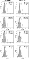
TSP1−/− retinal PCs expressed reduced levels of αvβ3-integrin. Expression levels of various integrins were determined by FACS analysis using specific antibodies as described in materials and methods. The shaded traces show staining in the absence of primary antibody. Please note the similar expression levels of β1-integrin, α6-integrin, and CD47 (integrin-associated protein, a TSP1 receptor) in TSP1+/+ and TSP1−/− PCs. Expression levels of αvβ3-integrin were significantly lower in TSP1−/− PCs (see text for quantifications). These experiments were repeated with three isolations of PCs with similar results.
Fig. 9.
Fig. 9.
Retinal PCs do not form an extensive cell network in Matrigel. The ability of TSP1+/+ and TSP1−/− retinal PCs to undergo capillary-like morphogenesis was evaluated by plating in Matrigel as described in materials and methods. Please note the minimal organization of retinal PCs cultured in Matrigel regardless of TSP1 status. These experiments were repeated with three isolations of PCs with similar results.
Fig. 10.
Fig. 10.
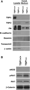
TSP1−/− retinal PCs exhibited altered production of ECM proteins and Akt1 activation. A: levels of various ECM proteins, including TSP1, TSP2, fibronectin (FN), and tenascin C, were determined by Western blot analysis of cell lysates and conditioned medium as described in materials and methods. TSP1+/+ PCs expressed significant amounts of TSP1, which was mainly cell associated, whereas TSP1−/− cells lacked TSP1. TSP1−/− PCs produced increased levels of TSP2 and FN but showed similar levels of tenascin C. β-Actin was used as a loading control for cell lysates. B: levels of Akt1 and its phosphorylated (active) form (pAkt), as well as its downstream target endothelial nitric oxide synthase (eNOS), were evaluated by Western blot analysis of cell lysates as described in materials and methods. Please note the increased levels of Akt1 and its phosphorylated (active) form in TSP1−/− compared with TSP1+/+ PCs. We were unable to detect eNOS expression in retinal PCs. These experiments were repeated with three isolations of PCs with similar results.

References

    1. Andrae J, Gallini R, Betsholtz C. Role of platelet-derived growth factors in physiology and medicine. Genes Dev 22: 1276–1312, 2008. - PMC - PubMed
    1. Buzney SM, Frank RN, Robison WG Jr. Retinal capillaries: proliferation of mural cells in vitro. Science 190: 985–986, 1975. - PubMed
    1. Buzney SM, Massicotte SJ, Hetu N, Zetter BR. Retinal vascular endothelial cells and pericytes. Differential growth characteristics in vitro. Invest Ophthalmol Vis Sci 24: 470–480, 1983. - PubMed
    1. Campochiaro PA Molecular targets for retinal vascular diseases. J Cell Physiol 210: 575–581, 2007. - PubMed
    1. Canfield AE, Sutton AB, Hoyland JA, Schor AM. Association of thrombospondin-1 with osteogenic differentiation of retinal pericytes in vitro. J Cell Sci 109: 343–353, 1996. - PubMed

Publication types

MeSH terms

LinkOut - more resources