Comparative Study
. 2009 Feb 4;29(5):1525-37.
doi: 10.1523/JNEUROSCI.5575-08.2009.

The serum response factor and a putative novel transcription factor regulate expression of the immediate-early gene Arc/Arg3.1 in neurons

Affiliations
Comparative Study

The serum response factor and a putative novel transcription factor regulate expression of the immediate-early gene Arc/Arg3.1 in neurons

Sean A Pintchovski et al. J Neurosci. .

Abstract

The immediate-early effector gene Arc/Arg3.1 is robustly upregulated by synaptic activity associated with learning and memory. Here we show in primary cortical neuron culture that diverse stimuli induce Arc expression through new transcription. Searching for regulatory regions important for Arc transcription, we found nine DNaseI-sensitive nucleosome-depleted sites at this genomic locus. A reporter gene encompassing these sites responded to synaptic activity in an NMDA receptor-dependent manner, consistent with endogenous Arc mRNA. Responsiveness mapped to two enhancer regions approximately 6.5 kb and approximately 1.4 kb upstream of Arc. We dissected these regions further and found that the proximal enhancer contains a functional and conserved "Zeste-like" response element that binds a putative novel nuclear protein in neurons. Therefore, activity regulates Arc transcription partly by a novel signaling pathway. We also found that the distal enhancer has a functional and highly conserved serum response element. This element binds serum response factor, which is recruited by synaptic activity to regulate Arc. Thus, Arc is the first target of serum response factor that functions at synapses to mediate plasticity.


Figures

Figure 1.
Figure 1.
Diverse stimuli induce new Arc transcription. A, Diagram of Arc pre-mRNA and the qfRT-PCR primer pairs (arrowheads) used to detect its levels. B, Induction of nascent Arc pre-mRNA was measured by qfRT-PCR after 6 h stimulations with either BDNF (100 ng/ml), the adenylyl cyclase activator forskolin (50 μm) and the phosphodiesterase inhibitor IBMX (100 μm), or 6 h after washout of chronic TTX (2 μm, 48 h; TTX pretreated). This induction was inhibited by APV (100 μm) added after washout. Values are mean ± SEM. N = 3 distinct cDNA pools, each amplified from a separate neuronal culture prep. Paired t test: **p < 0.01, *p < 0.05 versus control; n.s., not statistically significant.
Figure 2.
Figure 2.
In silico identification of putative regulatory regions containing conserved transcription factor binding sites. The online UCSC BLAT and the online EVOPRINTER analysis tools were used to identify seven upstream (A–G) and two downstream (H, I) regions (black rectangles) flanking Arc (gray rectangles) that are enriched in evolutionarily conserved sequences. Nucleotides within region B and regions F/G that are conserved in both sequence and relative genomic position are shown in black capital letters. In contrast, a portion of the genomic region between regions C and D demonstrates the relative lack of such conservation in the surrounding areas (see supplemental Fig. 3, available at www.jneurosci.org as supplemental material, for additional analysis). TESS was used to screen these conserved sequence elements for consensus binding sites, revealing a putative SRE (boxed) and an adjacent putative Ets/Elk-1 element (underlined) in region B as well as two putative Zeste-like binding sites (boxed) in regions F/G. All distances are from the Arc TSS.
Figure 3.
Figure 3.
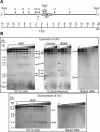
DHAs identify nucleosome-depleted regions flanking Arc. A, Probes used to map hypersensitive sites at the Arc locus. Genomic DNA fragments were detected by Southern blot probes that annealed either immediately upstream (“U”; black bar) or downstream (“D”; black bar) of the SspI restriction site in Arc. Arrows indicate the relative positions of the nine DNaseI HSs (A–I) identified at the Arc locus; gray circles represent DNaseI-inaccessible nucleosome-packaged regions. B, Top, DHAs identify seven discrete open regions (bands A–G; arrows) upstream of Arc. Some are present in both rat cortical neurons (RCNs) and PC12 cells (bands A, B, D, F, G); others are visible only in RCNs (band E) or only in PC12 cells (band C). There is a larger open smear (asterisks) at the TSS. The “naked” DNA negative control, performed using both PC12 and RCN DNA, exhibits only bands A and G, suggesting that they are either DHA artifacts or atypical sites and that the remaining bands and smear reflect the native chromatin structure upstream of Arc. Bottom, DHAs identify two discrete open regions (bands H, I) downstream of Arc in PC12 cells, but not in RCNs (data not shown). The naked DNA control demonstrates that both bands represent native chromatin structure. Blot contrasts were chosen to best represent all observed bands. Bands D and E (arrows and arrowheads) are more clearly visible in higher-contrast images (supplemental Fig. 2, available at www.jneurosci.org as supplemental material). All Southern blot images are representative of at least three experiments. Distances are in kb from the Arc TSS. The DNaseI concentration gradient, indicated above each blot, increases from left to right and includes a “no DNaseI” negative control in lane “O” (see Materials and Methods) (supplemental Fig. 1, available at www.jneurosci.org as supplemental material). PB: primary band resulting from SspI restriction cuts.
Figure 4.
Figure 4.
Stimulus-inducible distal and proximal enhancer regions. A, A strong correlation was observed between the genomic positions of the DHA HSs and the evolutionarily conserved regions, suggesting potential involvement in Arc transcription. A reporter with a 2.6-kb genomic fragment containing the proximal Arc promoter, which already contained four of the nine putative regulatory sites (D–G), was supplemented with the remaining upstream (A–C) and downstream (H, I) HSs. B, The new full-length Arc reporter is induced by synaptic activity. In agreement with activity-regulated expression of endogenous Arc mRNA, this reporter response is completely blocked by the NMDAR inhibitor APV or the ERK activation inhibitor UO126. C, A systematic deletional analysis of the new full-length Arc reporter. Left, “‡” indicates the reporter construct with the minimal deletions (ΔB and ΔF/G together) that result in a reduced response to synaptic activity. “Proximal” indicates the ∼900-bp basal promoter region immediately upstream of Arc. All values are percentages (mean ± SEM) of the maximum measurement for reporter ABCDEFG. N = 4, one-way ANOVA and post hoc Tukey's t tests: ***p < 0.001, *p < 0.05; n.s., not statistically significant. Right, “‡” indicates the reporter constructs with the minimal deletions (ΔB or ΔF/G individually) that result in a reduced stimulus-mediated response. This side-by-side construct comparison highlights how these two regions differentially contribute to BDNF- and forskolin-induced reporter activity. All values are percentages (mean ± SEM) of the maximum BDNF measurement for reporter ABCDEFG. N = 5, one-way ANOVA and post hoc Tukey's t tests: ***p < 0.001, **p < 0.01, *p < 0.05.
Figure 5.
Figure 5.
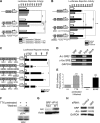
SRF mediates enhancer activity of distal region B. A, Region B is sufficient to enhance SV40 promoter activity in response to synaptic activity, and inactivating point mutations of SRE1 (mSRE1, with mutated base pairs underlined) reduce this response by ∼50%. This enhancer activity is completely blocked by the NMDAR inhibitor APV or the PKA inhibitor H89. All values are percentages (mean ± SEM) of the maximum induction for B. N = 4, one-way ANOVA and post hoc Tukey's t tests: ***p < 0.001. B, Region B is sufficient to enhance SV40 promoter activity in response to both BDNF and forskolin (consistent with Fig. 4C, right). SRE1 is necessary to mediate the response to BDNF but is not important for the response to forskolin. In contrast, region D has no effect on BDNF- or forskolin-induced SV40 promoter activity. All values are percentages (mean ± SEM) of the maximum for B. N = 5, one-way ANOVA and post hoc Tukey's t tests: ***p < 0.001, **p < 0.01; n.s., not statistically significant. C, Three putative SREs identified in silico were systematically inactivated by subtle point mutations (mSRE) in the context of the full-length Arc reporter. SRE1, located within region B, mediates ∼20% of the response to BDNF and is the only functional element identified. All values are percentages (mean ± SEM) of the maximum for ABCDEFG (top construct). N = 3, one-way ANOVA and post hoc Tukey's t tests: ***p < 0.001, **p < 0.01. D, ChIPs demonstrate that SRF physically associates in vivo with SRE1. Semi-quantitative PCR shows that an SRF-specific antibody enriches for genomic region B compared with a nonspecific control IgG. “Input” refers to the genomic input DNA used for the immunoprecipitation. “Mock” refers to a no-template control PCR. Gel images are representative of three experiments. E, Stimulation paradigms that induce Arc also activate SRF. BDNF treatment or chronic TTX pretreatment and washout induce SRE-Luciferase reporter gene activity, indicating that SRF mediates transcriptional responses to both stimuli. Values are mean ± SEM. N = 2, unpaired t test: ***p < 0.001. F, Synaptic activity induced by chronic TTX washout (“Washed”) leads to the phosphorylation of ERK1/2 (“pERK”), an upstream regulator of SRF-containing SRE-bound complexes. Total ERK1/2 expression levels are unaffected (“ERK”). All Western blot images are representative of three experiments. G, Constitutively active SRF (SRF-VP16) is sufficient to induce endogenous Arc protein expression. Wild-type SRF-VP16 (“WT”) robustly induces Arc expression, while a SRF-VP16 mutant (“Mut”) lacking the SRE DNA-binding domain of SRF does not. Actin served as a loading control. All Western blot images are representative of two experiments. H, SRF is important for the expression of endogenous Arc. Specific knockdown of SRF expression causes a corresponding decrease in BDNF-induced Arc levels, compared with a negative control siRNA that has no effect on SRF levels or BDNF-induced Arc expression. GAPDH served as a loading control. All Western blot images are representative of two biological experiments performed in triplicate.
Figure 6.
Figure 6.
A putative novel factor regulates proximal enhancer regions F/G. A, Regions F/G are sufficient to enhance SV40 promoter activity in response to synaptic activity. This enhancer activity is blocked by the NMDAR inhibitor APV or the PKA inhibitor H89. Inactivating point mutations of Zeste1 (mZ1, with mutated base pairs underlined) demonstrate that this site is necessary to mediate this enhanced response. All values are percentages (mean ± SEM) of the maximum induction for regions F/G. N = 3, one-way ANOVA and post hoc Tukey's t tests: ***p < 0.001, **p < 0.01, *p < 0.05; n.s., not statistically significant. B, Regions F/G are sufficient to enhance SV40 promoter activity in response to BDNF but not to forskolin (consistent with Fig. 4C, right). Inactivating point mutations of Zeste1 demonstrate that this site is necessary to mediate the enhanced response to BDNF. Deletion of an 86-bp stretch containing the Zeste1 and Zeste2 elements (Δ86bp) reduces BDNF-induced SV40 promoter activity below baseline, suggesting the presence of possible repressor elements in the remaining region. Region A also suppresses BDNF-induced SV40 promoter activity. All values are percentages (mean ± SEM) of the maximum induction for regions F/G. N = 5, one-way ANOVA and post hoc Tukey's t tests: ***p < 0.001; n.s., not statistically significant. C, EMSAs demonstrate that a nuclear protein factor or factors physically associate with the Zeste1 element. Several distinct mobility shifts (arrows) are observed when the wild-type Zeste1 probe (Z1) is pre-incubated with nuclear protein extracts (“Extract”), while the mutant Zeste1 probe (mZ1, containing the same point mutations that eliminate enhancer activity) did not show these corresponding shifts. The single shift observed for mZ1 (indicated by an asterisk) might represent a nonspecific binding event. Blot images are representative of three experiments.
Figure 7.
Figure 7.
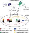
A model of Arc transcriptional regulation, showing how the activity-dependent PKA and ERK signaling pathways contribute to Arc transcription. Synaptic activity drives Arc transcription by inducing SRF/TCF activity in an NMDAR-, PKA-, and ERK-dependent manner (Figs. 4B, 5A, 5B, 5E–H). SRF binds to a conserved SRE in distal open region B (Figs. 3, 5D), and a conserved TCF binding site is found immediately downstream (Fig. 2). An unidentified enhancer factor likely also binds to region B to mediate the remaining response to synaptic activity and PKA (Figs. 5A, 5B). Synaptic activity also induces Arc transcription through a Zeste-like response element (ZRE) in proximal open regions F/G (Figs. 3, 6C) in an NMDAR-, PKA-, and ERK-dependent manner (Figs. 4B, 6A, 6B). Tyrosine receptor kinase B (TrkB) activation by BDNF induces Arc transcription via SRE and ZRE in an ERK-dependent manner (Figs. 4B, 5B, 6B). Some unidentified repressor factors likely bind to region A (Figs. 4C, 6B) and to regions F/G (Fig. 6B) to regulate stimulus-induced inhibition of reporter activity. The role of these putative repressors in Arc expression remains unclear.

Similar articles

Cited by

References

    1. Biggin MD, Bickel S, Benson M, Pirrotta V, Tjian R. Zeste encodes a sequence-specific transcription factor that activates the Ultrabithorax promoter in vitro. Cell. 1988;53:713–722. - PubMed
    1. Blaud M, Vossen C, Joseph G, Alazard R, Erard M, Nieto L. Characteristic patterns of N Oct-3 binding to a set of neuronal promoters. J Mol Biol. 2004;339:1049–1058. - PubMed
    1. Bode J, Goetze S, Heng H, Krawetz SA, Benham C. From DNA structure to gene expression: mediators of nuclear compartmentalization and dynamics. Chromosome Res. 2003;11:435–445. - PubMed
    1. Buchwalter G, Gross C, Wasylyk B. Ets ternary complex transcription factors. Gene. 2004;324:1–14. - PubMed
    1. Chen JD, Pirrotta V. Multimerization of the Drosophila zeste protein is required for efficient DNA binding. EMBO J. 1993;12:2075–2083. - PMC - PubMed

Publication types

MeSH terms

Substances

LinkOut - more resources