Comparative Study
. 2009 Feb 4;29(5):1544-53.
doi: 10.1523/JNEUROSCI.4491-08.2009.

Positron emission tomography imaging demonstrates correlation between behavioral recovery and correction of dopamine neurotransmission after gene therapy

Affiliations
Comparative Study

Positron emission tomography imaging demonstrates correlation between behavioral recovery and correction of dopamine neurotransmission after gene therapy

Ludovic Leriche et al. J Neurosci. .

Abstract

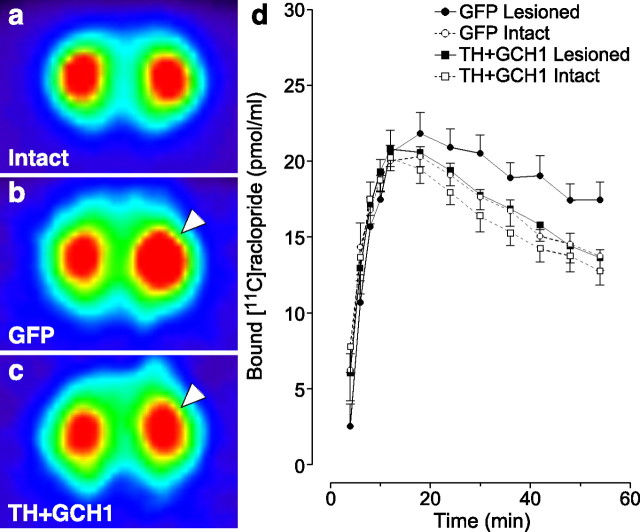
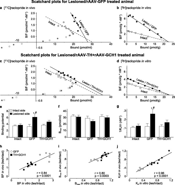
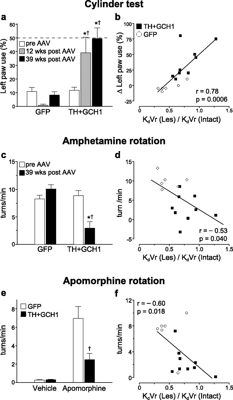
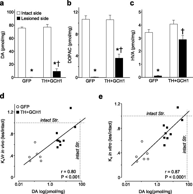
In vivo gene transfer using viral vectors is an emerging therapy for neurodegenerative diseases with a clinical impact recently demonstrated in Parkinson's disease patients. Recombinant adeno-associated viral (rAAV) vectors, in particular, provide an excellent tool for long-term expression of therapeutic genes in the brain. Here we used the [(11)C]raclopride [(S)-(-)-3,5-dichloro-N-((1-ethyl-2-pyrrolidinyl)methyl)-2-hydroxy-6-methoxybenzamide] micro-positron emission tomography (PET) technique to demonstrate that delivery of the tyrosine hydroxylase (TH) and GTP cyclohydrolase 1 (GCH1) enzymes using an rAAV5 vector normalizes the increased [(11)C]raclopride binding in hemiparkinsonian rats. Importantly, we show in vivo by microPET imaging and postmortem by classical binding assays performed in the very same animals that the changes in [(11)C]raclopride after viral vector-based enzyme replacement therapy is attributable to a decrease in the affinity of the tracer binding to the D(2) receptors, providing evidence for reconstitution of a functional pool of endogenous dopamine in the striatum. Moreover, the extent of the normalization in this non-invasive imaging measure was highly correlated with the functional recovery in motor behavior. The PET imaging protocol used in this study is fully adaptable to humans and thus can serve as an in vivo imaging technique to follow TH + GCH1 gene therapy in PD patients and provide an additional objective measure to a potential clinical trial using rAAV vectors to deliver l-3,4-dihydroxyphenylanaline in the brain.


Figures

Figure 1.
Figure 1.
Striatal gene transfer of TH and GCH1 genes using rAAV5 vectors. The efficiency of the rAAV5–TH vector was assessed by immunohistochemical staining of sections with antibodies against TH (a, d) and GCH1 (b, e) proteins as well as the NeuN neuronal marker (c, f). d–f are high-power views from the same sections as illustrated in a–c, respectively. TH staining shows a widespread expression of the transgenic human TH protein, in which numerous cell bodies and their dendritic projections are filled in the dorsal striatum (d). GCH1 expression is visualized using an antibody that specifically recognized the human GCH1 protein (b, e). Nonspecific effects of transduction was assessed by NeuN staining, which indicated that there were no apparent toxicity of striatal neurons on the injected side of the brain (c, f). Coexpression of TH and GCH1 proteins restored the in vivo DOPA synthesis capacity to normal levels in the lesioned striatum (g). * indicates different from the intact side; † indicates different from the respective lesion side from the TH + GCH1 group. Kruskal–Wallis nonparametric test, χ2(3,19) = 12.23, p < 0.01, followed by post hoc comparisons using Mann–Whitney U test with Bonferroni's correction. Scale bar: a–c, 500 μm; d–f, 25 μm.
Figure 2.
Figure 2.
[11C]raclopride PET experiments. The FORE-OSEM algorithm reconstructed tomographic data are illustrated as pseudocolored images indicating radioactivity levels high to low (from red–yellow to green–blue), in an intact rat (a), in a lesioned rAAV5–GFP-injected control rat (b), and in a lesioned rat treated with the TH + GCH1 therapeutic genes (c). Note the large increase on the [11C]raclopride signal on the lesion side (right hemisphere as indicated with arrowhead) in b and the normalization of the binding in the treated animal (arrowhead) in c. The kinetics of specific [11C]raclopride binding is plotted against time to illustrate the increased signal on the lesion side of the rAAV5–GFP-injected control animals starting at 24 min and maintained for the rest of the scanning period, whereas in the rAAV5–TH + rAAV5–GCH1-treated rats, similar levels of binding were seen between the two sides (d).
Figure 3.
Figure 3.
Quantification of the [11C]raclopride PET data. Analysis of the raclopride striatal binding was conducted by plotting the binding data in Scatchard plots both in vivo (a, c) and in vitro (b, d). The data points from the lesion/GFP side clearly deviated from those on the intact side (a, b), whereas the lesion/TH + GCH1 binding mimicked the binding of the intact side (c, d). Analysis of the data from in vivo [11C]raclopride PET scans expressed as BP to the striatal D2 DA receptors indicated a significant increase on the lesion side of the GFP-injected hemiparkinsonian rats and restoration of BP values to control values in the lesion side of the TH + GCH1-injected animals (e). To resolve the precise source of the change in BP, we calculated the apparent D2 DA receptor density (Bmax; f) and affinity of the binding to the receptors (KdVr − apparent in vivo Kd; g) by calculating the x-intercept and slope of the best-fit line from the quantitative PET dataset at the equilibrium state (12–50 min after injection; black symbols) and found that the changes were essentially attributable to a decrease in the KdVr and not an increase of the Bmax values. The in vivo estimates of BP, Bmax, and KdVr were tightly correlated to the corresponding in vitro values from the same animals (h–j). Statistics in e: two-way ANOVA, χ2(5,51) = 18.88, p < 0.005; g: two-way ANOVA, χ2(5,51) = 18.70, p < 0.005. The two-way analysis was followed by individual contrasts with Bonferroni's correction in both tests. Regression line fits in h–j are two-tailed significance of the Pearson's product-moment correlation coefficient. * indicates different from intact side; # indicates different from normal control.
Figure 4.
Figure 4.
Correlation between the in vivo PET data and the motor behavioral performance. The animals were tested on spontaneous motor behaviors using the cylinder test (a), amphetamine (c), and apomorphine (e) induced rotations. The use of the affected paw (left side) in the cylinder test was severely impaired after lesion. This impairment was maintained in rAAV5–GFP-injected animals, whereas the treatment group showed a significant recovery of function (a). The improvement in the cylinder test, expressed as difference between the pre-vector and post-vector injection time points, was highly correlated with the KdVr values derived from the striatal PET data (b). The correlations between drug-induced rotation and KdVr values were weaker (d, f. Statistics in a: two-way ANOVA, χ2(5,44) = 31.68, p < 0.001; c, two-way ANOVA, χ2(3,29) = 23.65, p < 0.001; e, two-way ANOVA, χ2(3,29) = 35.60, p < 0.001. All two-way analysis was followed by individual contrasts with Bonferroni's correction. Regression line fits in b, d, and f are two-tailed significance of the Pearson's product-moment correlation coefficient. * indicates different from pre-transduction score; † indicates different from lesion/GFP.
Figure 5.
Figure 5.
Tissue DA, DOPAC, and HVA levels were determined by HPLC (a–c). There was a significant restoration of all three metabolites in the rAAV5–TH + rAAV5–GCH1-treated rats. The tissue DA levels were well correlated with both in vivo KdVr (d) and in vitro Kd (e) values. Kruskal–Wallis nonparametric test, χ2(3,29) = 24.52 (DA), χ2(3,29) = 23.19 (DOPAC), and χ2(3,29) = 17.56 (HVA), p < 0.001 for all comparisons, followed by post hoc comparisons using Mann–Whitney U test with Bonferroni's correction. * indicates different from intact side; † indicates different from lesion/GFP.

References

    1. Antonini A, Leenders KL, Vontobel P, Maguire RP, Missimer J, Psylla M, Günther I. Complementary PET studies of striatal neuronal function in the differential diagnosis between multiple system atrophy and Parkinson's disease. Brain. 1997;120:2187–2195. - PubMed
    1. Carlsson T, Winkler C, Burger C, Muzyczka N, Mandel RJ, Cenci A, Björklund A, Kirik D. Reversal of dyskinesias in an animal model of Parkinson's disease by continuous l-DOPA delivery using rAAV vectors. Brain. 2005;128:559–569. - PubMed
    1. Carlsson T, Björklund T, Kirik D. Restoration of the striatal dopamine synthesis for Parkinson's disease: viral vector-mediated enzyme replacement strategy. Curr Gene Ther. 2007;7:109–120. - PubMed
    1. Chefer SI, Kimes AS, Matochik JA, Horti AG, Kurian V, Shumway D, Domino EF, London ED, Mukhin AG. Estimation of D2-like receptor occupancy by dopamine in the putamen of hemiparkinsonian monkeys. Neuropsychopharmacology. 2008;33:270–278. - PubMed
    1. Chritin M, Savasta M, Mennicken F, Bal A, Abrous DN, Le Moal M, Feuerstein C, Herman JP. Intrastriatal dopamine-rich implants reverse the increase of dopamine D2 receptor mRNA levels caused by lesion of the nigrostriatal pathway: a quantitative in situ hybridization study. Eur J Neurosci. 1992;4:663–672. - PubMed

Publication types

MeSH terms