

The first structure of a cold-adapted superoxide dismutase (SOD): biochemical and structural characterization of iron SOD from Aliivibrio salmonicida
- PMID: 19193992
- PMCID: PMC2635881
- DOI: 10.1107/S1744309109001110
The first structure of a cold-adapted superoxide dismutase (SOD): biochemical and structural characterization of iron SOD from Aliivibrio salmonicida
Abstract
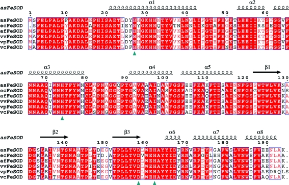
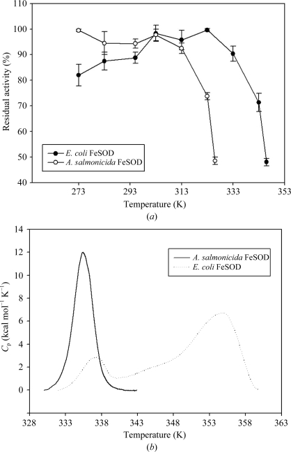
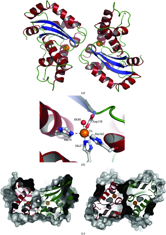
Superoxide dismutases (SODs) are metalloenzymes that catalyse the dismutation of the superoxide radical anion into O(2) and H(2)O(2) in a two-step reaction. The crystal structure of the iron superoxide dismutase from the cold-adapted and fish-pathogenic bacterium Aliivibrio salmonicida (asFeSOD) has been determined and refined to 1.7 A resolution. The protein has been characterized and compared with the closely related homologous iron superoxide dismutase from the mesophilic Escherichia coli (ecFeSOD) in an attempt to rationalize its environmental adaptation. ecFeSOD shares 75% identity with asFeSOD. Compared with the mesophilic FeSOD, the psychrophilic FeSOD has distinct temperature differences in residual activity and thermostability that do not seem to be related to structural differences such as intramolecular or intermolecular ion bonds, hydrogen bonds or cavity sizes. However, an increased net negative charge on the surface of asFeSOD may explain its lower thermostability compared with ecFeSOD. Activity measurements and differential scanning calorimetry measurements revealed that the psychrophilic asFeSOD had a thermostability that was significantly higher than the optimal growth temperature of the host organism.
Figures
Similar articles
-
Structure and flexibility in cold-adapted iron superoxide dismutases: the case of the enzyme isolated from Pseudoalteromonas haloplanktis.J Struct Biol. 2010 Dec;172(3):343-52. doi: 10.1016/j.jsb.2010.08.008. Epub 2010 Aug 21. J Struct Biol. 2010. PMID: 20732427
-
Comparison of protein-water interactions in psychrophilic, mesophilic, and thermophilic Fe-SOD.Protein Pept Lett. 2014 Jun;21(6):578-83. doi: 10.2174/0929866521666140108110050. Protein Pept Lett. 2014. PMID: 24410726
-
Thermostability of manganese- and iron-superoxide dismutases from Escherichia coli is determined by the characteristic position of a glutamine residue.Eur J Biochem. 2002 Nov;269(21):5137-48. doi: 10.1046/j.1432-1033.2002.03200.x. Eur J Biochem. 2002. PMID: 12392545
-
Roles of manganese and iron in the regulation of the biosynthesis of manganese-superoxide dismutase in Escherichia coli.FEMS Microbiol Rev. 1994 Aug;14(4):315-23. doi: 10.1111/j.1574-6976.1994.tb00105.x. FEMS Microbiol Rev. 1994. PMID: 7917419 Review.
-
Characterization of two copper/zinc superoxide dismutases (Cu/Zn-SODs) from the desert beetle Microdera punctipennis and their activities in protecting E. coli cells against cold.Cryobiology. 2019 Apr;87:15-27. doi: 10.1016/j.cryobiol.2019.03.006. Epub 2019 Mar 16. Cryobiology. 2019. PMID: 30890324 Review.
Cited by
-
Psychrophily and catalysis.Biology (Basel). 2013 Apr 16;2(2):719-41. doi: 10.3390/biology2020719. Biology (Basel). 2013. PMID: 24832805 Free PMC article.
-
Crystal structure of an iron superoxide dismutase from the pathogenic amoeba Acanthamoeba castellanii.Acta Crystallogr F Struct Biol Commun. 2019 Jul 1;75(Pt 7):480-488. doi: 10.1107/S2053230X19008112. Epub 2019 Jun 26. Acta Crystallogr F Struct Biol Commun. 2019. PMID: 31282867 Free PMC article.
-
Tolerance analysis of chloroplast OsCu/Zn-SOD overexpressing rice under NaCl and NaHCO3 stress.PLoS One. 2017 Oct 11;12(10):e0186052. doi: 10.1371/journal.pone.0186052. eCollection 2017. PLoS One. 2017. PMID: 29020034 Free PMC article.
-
Herbicides Tolerance in a Pseudomonas Strain Is Associated With Metabolic Plasticity of Antioxidative Enzymes Regardless of Selection.Front Microbiol. 2021 Jun 22;12:673211. doi: 10.3389/fmicb.2021.673211. eCollection 2021. Front Microbiol. 2021. PMID: 34239509 Free PMC article.
-
Regulation of Mitochondrial Dynamics in Parkinson's Disease-Is 2-Methoxyestradiol a Missing Piece?Antioxidants (Basel). 2021 Feb 6;10(2):248. doi: 10.3390/antiox10020248. Antioxidants (Basel). 2021. PMID: 33562035 Free PMC article. Review.
References
-
- Aghajari, N., Feller, G., Gerday, C. & Haser, R. (1998). Structure, 6, 1503–1516. - PubMed
-
- Amano, A., Shizukuishi, S., Tsunemitsu, A. & Tsunasawa, S. (1992). Oral Microbiol. Immunol.7, 368–371. - PubMed
-
- Barondeau, D. P., Kassmann, C. J., Bruns, C. K., Tainer, J. A. & Getzoff, E. D. (2004). Biochemistry, 43, 8038–8047. - PubMed
-
- Benov, L. T. & Fridovich, I. (1994). J. Biol. Chem.269, 25310–25314. - PubMed
-
- Bentahir, M., Feller, G., Aittaleb, M., Lamotte-Brasseur, J., Himri, T., Chessa, J. P. & Gerday, C. (2000). J. Biol. Chem.275, 11147–11153. - PubMed
Publication types
MeSH terms
Substances
LinkOut - more resources
Full Text Sources
Medical
