

Expression, purification, crystallization and preliminary X-ray diffraction analysis of the transcriptional repressor SirR from Mycobacterium tuberculosis H37Rv
- PMID: 19194009
- PMCID: PMC2635875
- DOI: 10.1107/S1744309108043534
Expression, purification, crystallization and preliminary X-ray diffraction analysis of the transcriptional repressor SirR from Mycobacterium tuberculosis H37Rv
Abstract
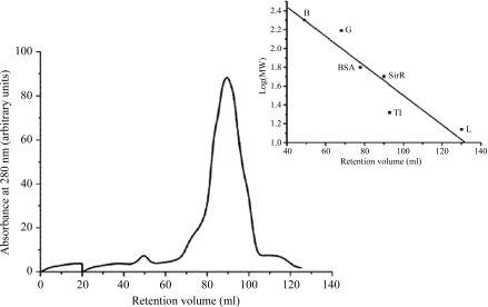
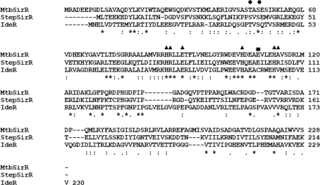
SirR, a metal-dependent transcriptional repressor from Mycobacterium tuberculosis (Rv2788), was cloned in pQE30 expression vector with an N-terminal His(6) tag for heterologous overexpression in Escherichia coli M15 (pREP4) cells and purified to homogeneity using chromatographic procedures. The purified protein was crystallized using the sitting-drop vapour-diffusion technique. The crystals belonged to the tetragonal space group P4(1)2(1)2/P4(3)2(1)2, with unit-cell parameters a = 105.21, b = 105.21, c = 144.85 A. The X-ray diffraction data were processed to a maximum resolution of 2.5 A. The Matthews coefficient suggests the presence of two (V(M) = 4.01 A(3) Da(-1)) to four (V(M) = 2.0 A(3) Da(-1)) molecules in the asymmetric unit. Calculation of the self-rotation function shows a crystallographic fourfold symmetry axis along the z axis (chi = 90 degrees) and also a twofold symmetry axis around the z axis (chi = 180 degrees).
Figures


Similar articles
-
Expression, purification and preliminary X-ray analysis of the C-terminal domain of an arginine repressor protein from Mycobacterium tuberculosis.Acta Crystallogr Sect F Struct Biol Cryst Commun. 2007 Nov 1;63(Pt 11):936-9. doi: 10.1107/S1744309107046374. Epub 2007 Oct 24. Acta Crystallogr Sect F Struct Biol Cryst Commun. 2007. PMID: 18007044 Free PMC article.
-
Crystallization and preliminary X-ray diffraction studies of Streptococcus pyogenes plasmid pSM19035-encoded omega transcriptional repressor.Acta Crystallogr D Biol Crystallogr. 1999 Dec;55(Pt 12):2041-2. doi: 10.1107/s0907444999012275. Acta Crystallogr D Biol Crystallogr. 1999. PMID: 10666585
-
Expression, purification, crystallization and preliminary X-ray diffraction studies of glyceraldehyde-3-phosphate dehydrogenase 1 from methicillin-resistant Staphylococcus aureus (MRSA252).Acta Crystallogr Sect F Struct Biol Cryst Commun. 2008 Oct 1;64(Pt 10):929-32. doi: 10.1107/S1744309108027504. Epub 2008 Sep 30. Acta Crystallogr Sect F Struct Biol Cryst Commun. 2008. PMID: 18931438 Free PMC article.
-
Cloning, overexpression, purification, crystallization and preliminary X-ray diffraction analysis of glyceraldehyde-3-phosphate dehydrogenase from Antheraea mylitta.Acta Crystallogr Sect F Struct Biol Cryst Commun. 2009 Sep 1;65(Pt 9):937-40. doi: 10.1107/S174430910903214X. Epub 2009 Aug 26. Acta Crystallogr Sect F Struct Biol Cryst Commun. 2009. PMID: 19724138 Free PMC article.
-
Expression, purification, crystallization and preliminary X-ray diffraction studies of triosephosphate isomerase from methicillin-resistant Staphylococcus aureus (MRSA252).Acta Crystallogr Sect F Struct Biol Cryst Commun. 2009 Apr 1;65(Pt 4):398-401. doi: 10.1107/S1744309109010112. Epub 2009 Mar 25. Acta Crystallogr Sect F Struct Biol Cryst Commun. 2009. PMID: 19342791 Free PMC article.
References
-
- Bradford, M. M. (1976). Anal. Biochem.72, 248–254. - PubMed
-
- Cole, S. T. et al. (1998). Nature (London), 393, 537–544. - PubMed
-
- Collaborative Computational Project, Number 4 (1994). Acta Cryst. D50, 760–763. - PubMed
-
- Feese, M. D., Ingason, B. P., Goranson-Siekierke, J., Holmes, R. K. & Hol, W. G. (2001). J. Biol. Chem.276, 5959–5965. - PubMed
-
- Gold, B., Rodriguez, G. M., Marras, S. A., Pentecost, M. & Smith, I. (2001). Mol. Microbiol.42, 851–865. - PubMed
Publication types
MeSH terms
Substances
LinkOut - more resources
Full Text Sources
Molecular Biology Databases
