. 2009 Aug;129(8):2011-21.
doi: 10.1038/jid.2008.458. Epub 2009 Feb 5.

Protein kinase Cepsilon inhibits UVR-induced expression of FADD, an adaptor protein, linked to both Fas- and TNFR1-mediated apoptosis

Affiliations

Protein kinase Cepsilon inhibits UVR-induced expression of FADD, an adaptor protein, linked to both Fas- and TNFR1-mediated apoptosis

Moammir Hasan Aziz et al. J Invest Dermatol. 2009 Aug.

Abstract

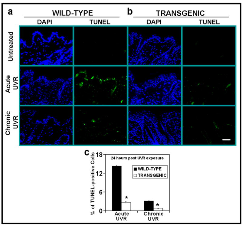
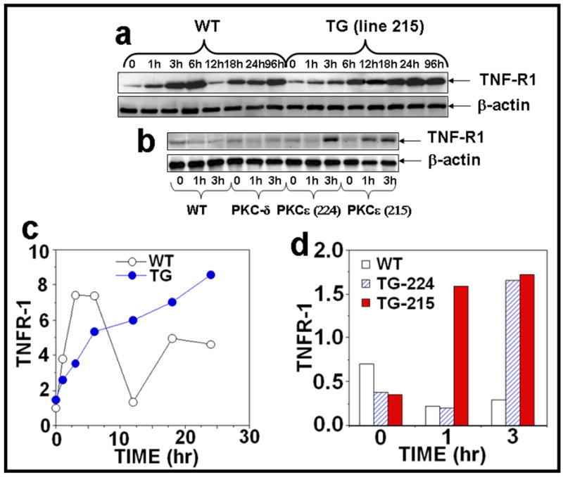
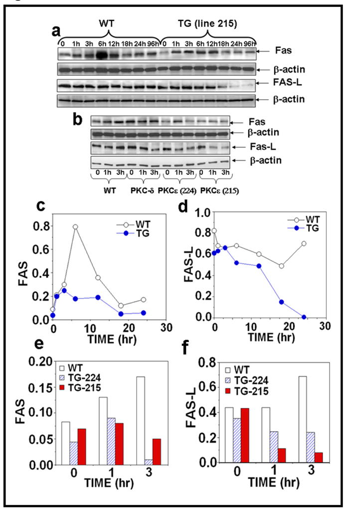
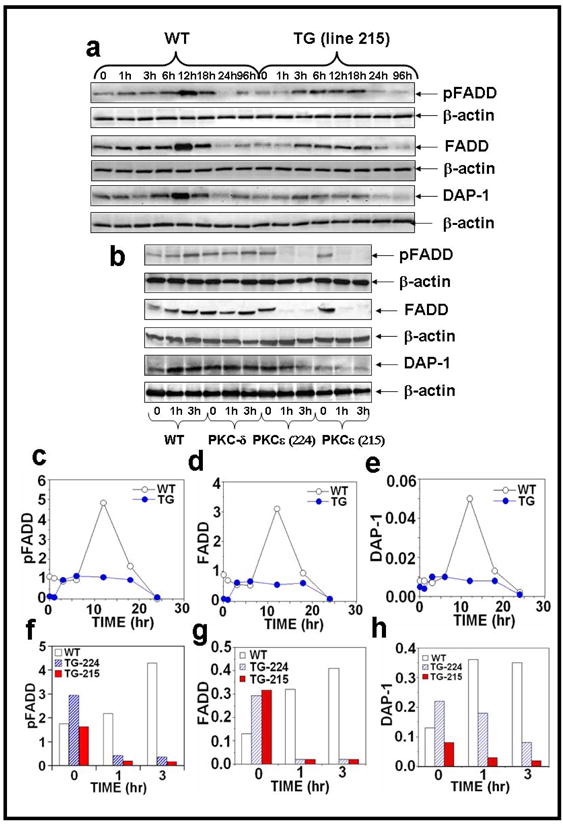
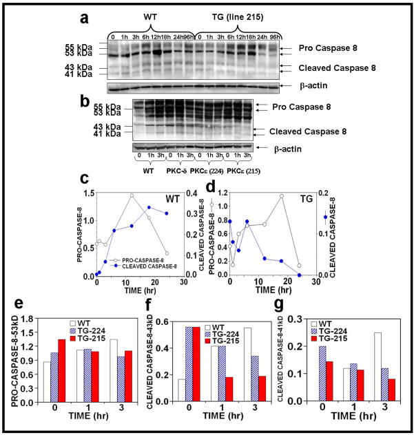
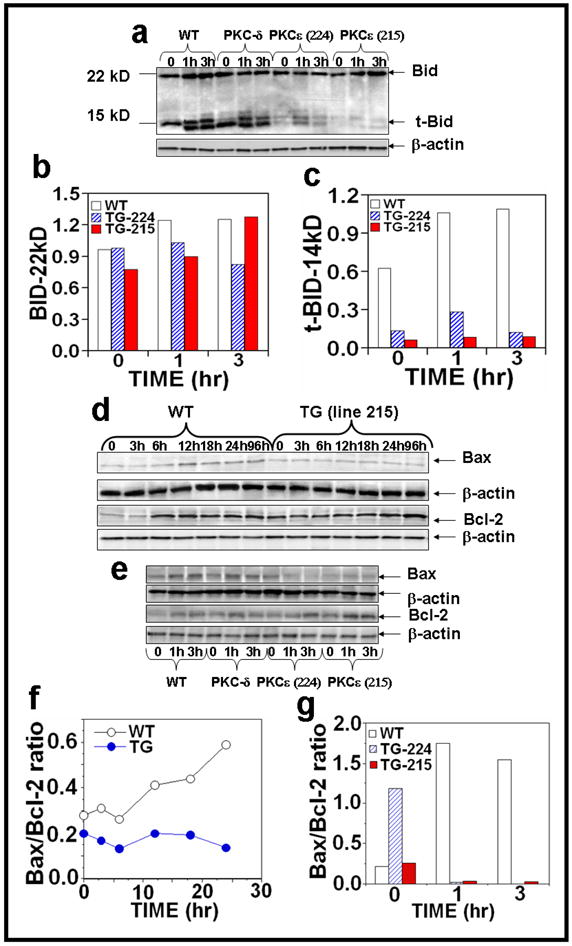
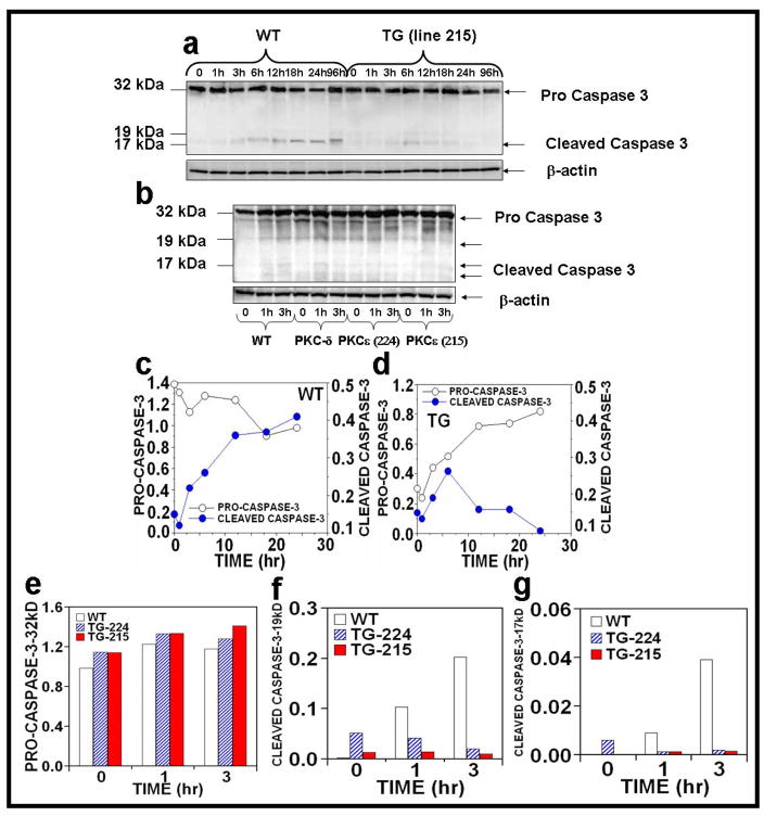
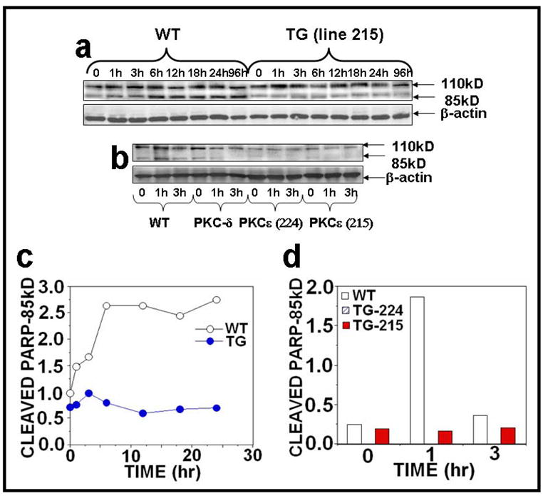
Protein kinase C (PKC)epsilon overexpression in FVB/N transgenic mice sensitized skin to UVR-induced development of squamous cell carcinomas and suppressed formation of sunburn cells, which are DNA-damaged keratinocytes undergoing apoptosis. Here, we elucidated the mechanisms associated with the inhibition of UVR-induced appearance of sunburn cells in PKCepsilon transgenic mice. We found that the inhibition of UVR-induced sunburn cell formation in PKCepsilon transgenic mice may be the result of the inhibition of the expression of Fas, Fas ligand, and the mammalian death adaptor protein termed Fas-associated with death domain (FADD). The adaptor protein FADD is the key component of the death-inducing signaling complex of both Fas and tumor necrosis factor receptor 1. A decreased expression of epidermal FADD was observed after a single UVR exposure. However, a complete loss of FADD expression was found after four (Monday, Wednesday, Friday, and Monday) repeated UVR exposures. FADD transmits apoptotic signals from death receptors to the downstream initiator caspase-8 and connects to the mitochondrial intrinsic apoptotic signal transduction pathway by the cleavage of Bid, a Bcl-2 family member. PKCepsilon-mediated loss of FADD expression inhibited UVR signals to the activation of both extrinsic and intrinsic apoptotic pathways.


Conflict of interest statement

CONFLICT OF INTEREST

None

Figures

Figure 1
Figure 1. PKCε overexpression suppresses UVR-induced apoptosis
PKCε transgenic mice (line 215) and their wild-type littermates were exposed to acute (single) UVR (4 kJ/m2) or chronic (repeated) UVR (2kJ/m2) four times (Monday, Wednesday, Friday, and Monday). The mice were sacrificed at 24 hours after last UVR exposure. Skin specimens were fixed in 10% neutral buffered formalin and embedded in paraffin. Skin sections of 4-μm thickness were cut and analyzed for the presence of apoptosis cells by TUNEL assay. (a) DAP1 counter stain and TUNNEL assay of WT mice at 0 and 24 hrs post acute or chronic UV exposures (b). DAP1 counter stain and TUNNEL assay of PKCε transgenic mice c: Quantitaion of TUNEL-positive cells 24 hours after acute or chronic UVR exposures. Four mice were sacrificed at each time point, and eight microscopic fields in each mouse were used to calculate average number of TUNEL-positive cells. *, p<0.005. Bars= 50 μm.
Figure 2
Figure 2. PKCε overexpression sensitizes skin to UVR-induced TNF-R1 expression
(a): PKCε transgenic mice (line 215) and wild-type littermates (4 mice per group) were exposed to single UVR (4 kJ/m2). The mice were sacrificed at 1, 3, 6, 12, 18, 24, and 96 hours after acute UVR exposure. (b): In a parallel experiment, PKCε (lines 215 and 224), PKCδ transgenic mice and wild-type littermates were exposed to UVR (2kJ/m2) four times (Monday, Wednesday, Friday, and Monday). The mice were sacrificed at 1 and 3 hours post fourth treatment of UVR. Mouse epidermal exrtracts were prepared and TNFR1 and β-actin were assessed by immunoblot analysis as described previously. (c,d): The quantification of proteins (normalized to β-actin).
Figure 3
Figure 3. PKCε overexpression suppresses UVR-induced Fas and Fas-L expression
Epidermal extract prepared for Fig. 2 was used to analyze Fas and Fas-L expression. Immunoblot analysis of Fas expression after (a) acute (b) chronic UVR exposed samples and (c–f) the quantification of Fas and Fas-L expression.
Figure 4
Figure 4. PKCε overexpression completely suppresses the level of pFADD, FADD and DAP-1 in UVR exposed PKCε transgenic mice
Epidermal extract prepared for Fig. 2 was used to analyze pFADD, FADD and DAP-1 expression. Immunoblot analysis of pFADD, FADD and DAP-1 expression after (a) acute and (b) chronic UVR exposures. (c–h): The quantification of pFADD, FADD and DAP-1 expression.
Figure 5
Figure 5. Effect of UVR on the activation of caspase-8 in UVR exposed PKCε transgenic mice
Epidermal extract prepared for Fig. 2 was used to analyze caspase 8 expression. Immunoblot analysis of caspase-8 expression after (a) acute (b) chronic UVR exposed samples. (c–g): The quantification of caspase-8 expression.
Figure 6
Figure 6. PKCε overexpression inhibits tBid and Bax/Bcl-2 ratio in UVR exposed PKCε transgenic mice
Epidermal extract prepared for Fig. 2 was used to analyze Bid and tBid expression and Bax and Bcl-2 expression after (a,e) chronic and (d) acute UVR exposure. Quantification of (b) Bid and (c) tBid and (f, g) Bax/Bcl-2 ratio after chronic UVR exposure.
Figure 7
Figure 7. Effect of UVR on the activation of caspase-3 in UVR exposed PKCε transgenic mice
Epidermal extract prepared for Fig. 2 was used to analyze caspase-3 expression. Immunoblot analysis of caspase-3 expression after (a) acute (b) chronic UVR exposure. (c–g): Quantification of caspase-3 expression.
Figure 8
Figure 8. PKCε overexpression inhibits PARP and its cleavage in UVR exposed PKCε transgenic mice
Epidermal extract prepared for Fig. 2 was used to analyze PARP and its cleavage. Immunoblot analysis of PARP and its cleavage product after (a) acute (b) chronic UVR exposure. (c and d): Quantification of PARP and its cleavage product.

Comment in

Similar articles

Cited by

References

    1. Anonymous skin cancer; American Cancer Society. Cancer Facts & Figures 2008. Atlanta: American Cancer Society; 2008.
    1. Kulms D, Düssmann H, Pöppelmann B, Ständer S, Schwarz A, Schwarz T. Apoptosis induced by disruption of the actin cytoskeleton is mediated via activation of CD95 (Fas/APO-1) Cell Death Differ. 2002;9:598–608. - PubMed
    1. Ashkenazi A, Dixit VM. Death receptors: signaling and modulation. Science. 1998;281:1305–8. - PubMed
    1. Balint EE, Vousden KH. Activation and activities of the p53 tumour suppressor protein. Br J Cancer. 2001;85:1813–23. - PMC - PubMed
    1. Baud V, Karin M. Signal transduction by tumor necrosis factor and its relatives. Trends Cell Biol. 2001;11:372–7. - PubMed

Publication types

MeSH terms

Substances