. 2009;4(2):e4352.
doi: 10.1371/journal.pone.0004352. Epub 2009 Feb 5.

At least ten genes define the imprinted Dlk1-Dio3 cluster on mouse chromosome 12qF1

Affiliations

At least ten genes define the imprinted Dlk1-Dio3 cluster on mouse chromosome 12qF1

John P Hagan et al. PLoS One. 2009.

Abstract

Background: Genomic imprinting is an exception to Mendelian genetics in that imprinted genes are expressed monoallelically, dependent on parental origin. In mammals, imprinted genes are critical in numerous developmental and physiological processes. Aberrant imprinted gene expression is implicated in several diseases including Prader-Willi/Angelman syndromes and cancer.

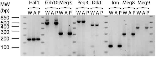
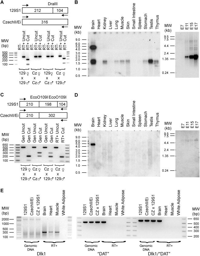
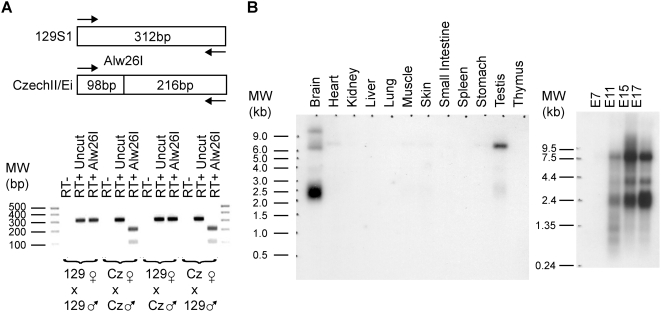
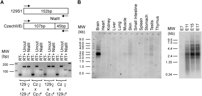
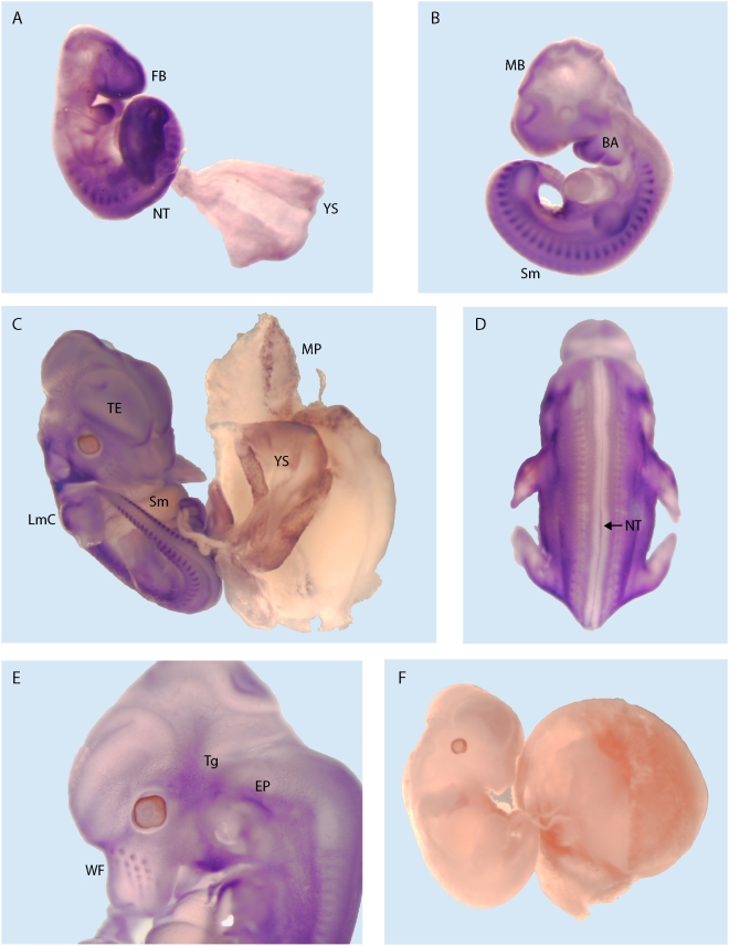
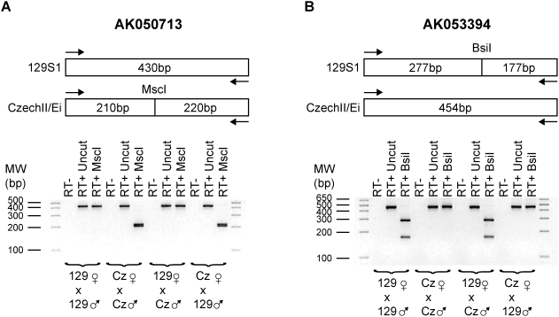
Methodology/principal findings: To identify novel imprinted genes, transcription profiling was performed on two uniparentally derived cell lines, androgenetic and parthenogenetic primary mouse embryonic fibroblasts. A maternally expressed transcript termed Imprinted RNA near Meg3/Gtl2 (Irm) was identified and its expression studied by Northern blotting and whole mounts in situ hybridization. The imprinted region that contains Irm has a parent of origin effect in three mammalian species, including the sheep callipyge locus. In mice and humans, both maternal and paternal uniparental disomies (UPD) cause embryonic growth and musculoskeletal abnormalities, indicating that both alleles likely express essential genes. To catalog all imprinted genes in this chromosomal region, twenty-five mouse mRNAs in a 1.96Mb span were investigated for allele specific expression.

Conclusions/significance: Ten imprinted genes were elucidated. The imprinting of three paternally expressed protein coding genes (Dlk1, Peg11, and Dio3) was confirmed. Seven noncoding RNAs (Meg3/Gtl2, Anti-Peg11, Meg8, Irm/"Rian", AK050713, AK053394, and Meg9/Mirg) are characterized by exclusive maternal expression. Intriguingly, the majority of these noncoding RNA genes contain microRNAs and/or snoRNAs within their introns, as do their human orthologs. Of the 52 identified microRNAs that map to this region, six are predicted to regulate negatively Dlk1, suggesting an additional mechanism for interactions between allelic gene products. Since several previous studies relied heavily on in silico analysis and RT-PCR, our findings from Northerns and cDNA cloning clarify the genomic organization of this region. Our results expand the number of maternally expressed noncoding RNAs whose loss may be responsible for the phenotypes associated with mouse pUPD12 and human pUPD14 syndromes.


Conflict of interest statement

Competing Interests: The authors have declared that no competing interests exist.

Figures

Figure 1
Figure 1. Androgenetic and Parthenogenetic PMEFs maintain appropriate imprinted gene expression.
RT-PCR was performed on indicated PMEF cell lines: W (Wild-type), A (Androgenetic), and P (Parthenogenetic). Histone acetyltransferase 1 (Hat1) is a biallelically expressed control. Dlk1 and Peg3 are controls for paternal expression while Grb10 and Meg3/Gtl2 are controls for maternal expression. With Irm primers 1307Up and 1581Dn, Irm RNA expression was undetectable in AG PMEF, while its expression was abundant in WT and PG PMEFs. Moreover, Meg8 and Meg9, two other genes in the large imprinted gene cluster that contains Irm, have expression patterns that are consistent with being imprinted with maternal expression. The PCR primers were Hat1 510Up/758Dn, Grb10 270Up/770Dn, Meg3 267Up/578Dn, Peg3 4430Up/4956Dn, Dlk1 830Up/1274Dn, Irm 988Up/1140Dn, Meg8 1Up/294Dn (AF498299) and Meg9 2614-3009.
Figure 2
Figure 2. Genomic Organization of Dlk1-Dio3 Imprinted Region.
This figure summarizes our results about the imprinting and structure of genes in this mouse region. Arrows denote transcriptional orientations. Genes written in black were shown to be biallelically expressed. Genes drawn in blue are paternally expressed, while those in red are maternally expressed. Genes written in yellow were undetectable by RT-PCR. For Begain transcribed from promoter 2, this transcript was coded blue as it is paternally expressed in sheep in a tissue specific manner, although in this study, RT-PCR failed to amplify a transcript derived from this promoter. Of these, accession numbers, AK141557, AK163826, AK048151, and AK044800, correspond to unspliced “transcripts” whose cDNA sequence ends in a polyA stretch of genomic DNA, suggesting that these “cDNAs” may be genomic DNA contamination in the Riken mouse cDNA libraries. For the bottom panel, exons are dark colored while introns are light. Purple lines represent the position of microRNA precursors. In all, fifty-two mouse microRNA precursors as listed in Table 2 map to this imprinted region.
Figure 3
Figure 3. Dlk1 is paternally expressed and has an abundant alternatively polyadenylated transcript in the mouse brain.
(A) A DraIII restriction site polymorphism between 129S1 and CzechII/Ei mice was utilized to determine that Dlk1 is paternally expressed. PCR primers were Dlk1 2Up/317Dn. (B) Northern analysis using the same Dlk1 PCR fragment as probe revealed that Dlk1 is widely expressed. Of the tissues investigated, only the adult brain is characterized by having an additional and abundant transcript that is roughly 4.5 kb in length. (C) Several ESTs cluster to the syntenic region of “DAT” (Dlk1 associated transcript). RT-PCR was done with “DAT” primers 1533Up/2044Dn. Using a EcoO109I restriction site polymorphism, “DAT” was shown to be like Dlk1 in its paternal expression. (D) Northern analysis on adult tissue samples of “DAT” detected a 4.5 kb transcript only in the brain. This band is identical in size to the long transcript of Dlk1 found in the brain. Moreover, the length of this transcript is greater than the distance (∼2.7 kb) between mouse Dlk1 polyA and “DAT” polyA. (E) RT-PCR was performed with various primer sets. Left most panel showed that Dlk1 is expressed in brain, heart and muscle. Middle panel shows that “DAT” is only detectable in brain and is consistent with Northern findings. For the right panel, PCR was performed with a primer set in which the upstream primer (Dlk1 1237Up) was before the canonical Dlk1 polyA site while the downstream primer was after this site (Dlk1 1805Dn). A band was detected only in the brain, indicating that “DAT” is an alternatively polyadenylated transcript of Dlk1. This result was confirmed by cDNA cloning (Accession numbers: EU434914-EU434917).
Figure 4
Figure 4. Meg3 is maternally expressed and highly expressed in the brain and testis.
(A) Alw26I restriction digests on RT-PCR products confirm that Meg3 is maternally expressed. For PCR, Meg3 267Up/578Dn primers were used. (B) Northern blots reveal that Meg3 is characterized by alternative splicing. In the adult mouse brain, the predominant band is roughly 2.4 kb in length while in testis is 6.5 kb.
Figure 5
Figure 5. Imprinting and expression analysis of mouse Peg11 and anti-Peg11.
(A) RT-PCR was performed on RNA derived from wild-type {W}, Androgenetic {A} and Parthenogenetic {P} PMEF cell lines. RT reactions were performed with either random hexamers or the strand-specific primers for Peg11 “477” or Anti-Peg11 “1052”. For PCR, Anti-Peg11 522Up/847Dn primers were used. These results demonstrate that in PMEFs Anti-Peg11 is expressed but not Peg11. (B) An NlaIII restriction site polymorphism between 129S1 and CzechII/Ei was utilized to demonstrate that Anti-Peg11 is maternally expressed in PMEFs. (C) As in panel A, three RT reactions were performed with the same PCR primers but on adult brain RNA derived from a mouse with a CzechII/Ei mother and 129S1 father. These results demonstrate that Anti-Peg11 and Peg11 are both expressed, although Anti-Peg11 is more abundant in mouse brain. Peg11 was paternally expressed, while Anti-Peg11 was maternally expressed. PCR on genomic DNA (Gen) revealed that the primers did not preferentially amplify either the 129S1 or CzechII/Ei alleles. (D) Northern for Peg11 and Anti-Peg11 using a double stranded DNA probe corresponding to Anti-Peg11 477-1052. Blue arrows indicate the position of Peg11 transcripts, while red arrows correspond to Anti-Peg11. This annotation was possible as the Northern was repeated with single stranded probes for both Peg11 and Anti-Peg11 (data not shown).
Figure 6
Figure 6. Meg8 is expressed from the maternal allele and highly in the brain and skin.
(A) The Meg8 alleles contain an informative restriction site polymorphism in that a HgaI site is present in the 129S1 allele in comparison to CzechII/Ei. RT-PCR products with AF498299 1Up/294Dn primers for Meg8 from the four intraspecific crosses were digested with and without HgaI, verifying the imprinted expression of this gene. (B) Northern blots were performed on adult tissues (left) and embryos harvested at different time points (right). Meg8 levels were highest in brain and testis. In addition, Meg8 RNA increases as a functional of embryonic development.
Figure 7
Figure 7. Irm is maternally expressed, alternatively spliced, and abundant in the adult brain.
(A). A single nucleotide polymorphism in Irm cDNA was identified at 1094A→T between 129S1 and CzechII/Ei mouse strains that creates a NlaIII restriction site in the CzechII/Ei Irm cDNA. RT-PCR was performed with Irm primers 988Up and 1140Dn. Restriction digests of RT-PCR products from intraspecific crosses between these two subspecies by NlaIII demonstrated that Irm is expressed in a monoallelic manner from the maternal allele. (B) An adult tissue (left) and total embryo from different gestational days (right) polyA+ RNA blot (left) was hybridized with a Irm/Rian exon 1 cDNA probe. A 2.5 kb transcript was predominantly detected in the brain and to a significantly lesser extent in testis, stomach, and muscle. Higher molecular weight bands of lower relative abundance in comparison to the 2.5 kb message were also seen that reflect the heterogeneity in spliced variants. Since Rian RNA is approximately 5.4 kb, the predominant RNA product from the Irm/Rian promoter is Irm RNA, while Rian accounts for at most 5% of total transcripts. This result is consistent with the relative abundance of ESTs that are specific for each of the alternatively spliced products.
Figure 8
Figure 8. Tissue distribution of the Irm transcripts in the mouse embryo revealed by in situ hybridization.
(A). On embryonic day 9.5 (E9.5), Irm expression is detected in the forebrain area (FB), first and second branchial arches (BA), in developing somites (Sm), and in the caudal neural tube (NT). A dispersed staining is observed in the extraembryonic tissues, particularly in the yolk sac (YS). (B). By E10.5 the brain expression domain shifts more in the midbrain area (MB) where the neuroepithelial lining shows the Irm staining. Similar to E9.5, expression persists both in the branchial arches and in the somites. (C). In E12 mouse conceptus, Irm transcripts could be also visualized in the forming limb cartilage (LmC), still clearly detectable in the telencephalon (TE) and caudal somites. The yolk sac (YS) and the maternal side of placenta (MP) are positive for Irm by in situ staining as well. The expression along the midline in the caudal neural tube (NT) is most likely associated with the floor plate area (D). In the head, prominent Irm transcript accumulation is observed in the trigeminal area (Tg), ear pinna (EP), and the developing whiskers hair follicle (WF, see panel E). (F). Hybridization with the sense Irm probe under the same conditions resulted in no specific signal.
Figure 9
Figure 9. AK050713 and AK053394 are maternally expressed.
(A) The AK050713 alleles contain an informative restriction site polymorphism in that a MscI site is present in the CzechII/Ei allele in comparison to 129S1. RT-PCR products with primers 784Up/1213Dn from the four intraspecific crosses were digested with and without MscI, verifying the imprinted expression of this gene. (B) Similarly, a BslI informative restriction site was utilized to demonstrate that AK053394 is maternally expressed using PCR primers 1647Up/2100Dn.
Figure 10
Figure 10. Meg9 transcripts are derived from the maternal allele and are widely expressed in adult tissues.
(A) Meg9 alleles between 129S1 and CzechII/Ei differ by a polymorphism that abolishes a MboII restriction site in the CzechII/Ei allele. Moreover, the CzechII/Ei allele harbors a fourteen nucleotide long insertion. RT-PCR (bottom) and MboII restriction digestion (top) of the Meg9 RT-PCR products demonstrated that this gene is maternally expressed. (B) Northern demonstrate that Meg9, like Dlk1 (see Figure 3B), is widely expressed in adult tissues and that the abundance of its transcripts increase as a function of embryonic age.
Figure 11
Figure 11. Allelic Expression of Dio3 and Anti-Dio3 (A) Dio3 is an atypical imprinted gene in that the expression occurs preferentially from the paternal allele.
Expression of the maternal allele occurs at roughly one-quarter the level of its paternal counterpart as evidenced by TaqI digests and (B) direct sequencing of RT-PCR products using Dio3 1216Up/1657Dn primers. (C) Anti-Dio3 is biallelically expressed as evidenced by MspI restriction digests and (D) direct sequencing of RT-PCR products using Dio3-AS 654Up/1197Dn primers.

Similar articles

Cited by

References

    1. McGrath J, Solter D. Completion of mouse embryogenesis requires both the maternal and paternal genomes. Cell. 1984;37:179–183. - PubMed
    1. Barton SC, Surani MA, Norris ML. Role of paternal and maternal genomes in mouse development. Nature. 1984;311:374–376. - PubMed
    1. Joyce JA, Schofield PN. Genomic imprinting and cancer. Mol Pathol. 1998;51:185–190. - PMC - PubMed
    1. Esteller M, Herman JG. Cancer as an epigenetic disease: DNA methylation and chromatin alterations in human tumours. J Pathol. 2002;196:1–7. - PubMed
    1. Ogawa O, Eccles MR, Szeto J, McNoe LA, Yun K, et al. Relaxation of insulin-like growth factor II gene imprinting implicated in Wilms' tumour. Nature. 1993;362:749–751. - PubMed

Publication types

MeSH terms