. 2009;4(2):e4376.
doi: 10.1371/journal.pone.0004376. Epub 2009 Feb 5.

A newly identified essential complex, Dre2-Tah18, controls mitochondria integrity and cell death after oxidative stress in yeast

Affiliations

A newly identified essential complex, Dre2-Tah18, controls mitochondria integrity and cell death after oxidative stress in yeast

Laurence Vernis et al. PLoS One. 2009.

Abstract

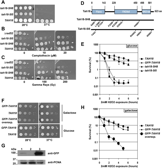
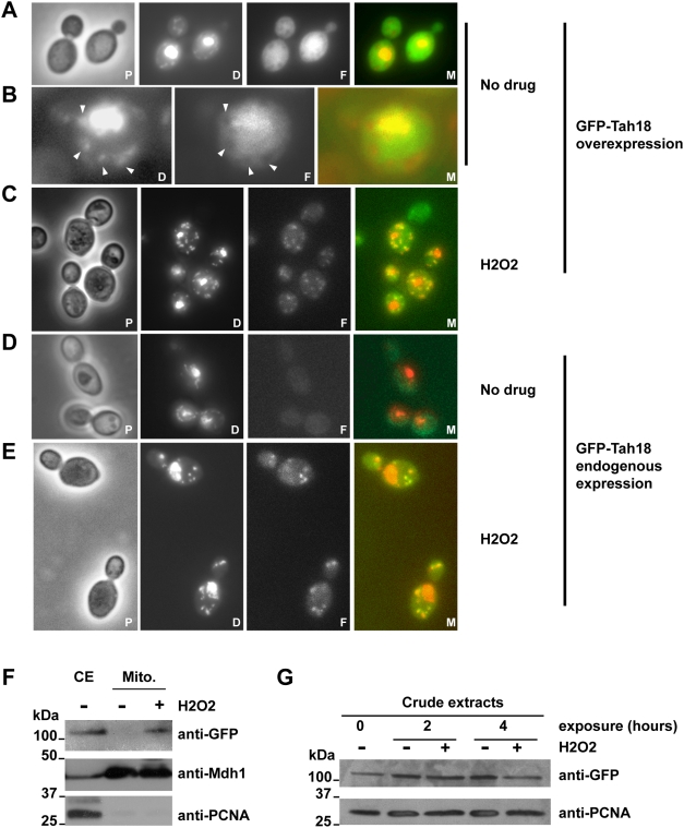
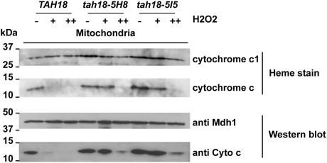
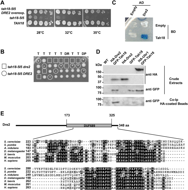
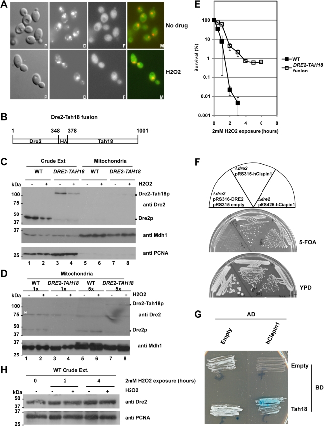
A mutated allele of the essential gene TAH18 was previously identified in our laboratory in a genetic screen for new proteins interacting with the DNA polymerase delta in yeast [1]. The present work shows that Tah18 plays a role in response to oxidative stress. After exposure to lethal doses of H(2)O(2), GFP-Tah18 relocalizes to the mitochondria and controls mitochondria integrity and cell death. Dre2, an essential Fe/S cluster protein and homologue of human anti-apoptotic Ciapin1, was identified as a molecular partner of Tah18 in the absence of stress. Moreover, Ciapin1 is able to replace yeast Dre2 in vivo and physically interacts with Tah18. Our results are in favour of an oxidative stress-induced cell death in yeast that involves mitochondria and is controlled by the newly identified Dre2-Tah18 complex.


Conflict of interest statement

Competing Interests: The authors have declared that no competing interests exist.

Figures

Figure 1
Figure 1. A–C. Tah18-5H8 and tah18-5I5 are thermosensitive mutants that are not involved in DNA repair after exposure to gamma rays or camptothecin.
Ten-fold dilutions of exponentially grown yeast cells were spotted onto YPD medium (A–B) or YPD containing Camptothecin as indicated (C). Gamma rays irradiation was performed using a 137Cs with a dose rate of 0.850 Gy/s. Growth was assessed after 3 days at 28°C unless indicated. A Δ rad52 mutant was used as a control. D. Tah18 exhibits three domains potentially involved in oxydo-reduction reactions. Flavodoxin, FAD-binding and NAD-binding domains were detected using Blast program. Mutations in tah18-5H8 and tah18-5I5 are indicated. E. Tah18-5H8 and tah18-5I5 are more resistant to H2O2 acute exposure than TAH18. Exponentially growing cells in YPD (1–5.106 cells per milliliter of culture) were exposed to 2 mM H2O2 during 1 to 6 hours before being plated onto YPD. After 3 days at 28°C, the number of colony forming units (CFU) was counted and expressed as a percentage of the number of CFU at time zero to assess survival rate. Points are means of three independent experiments. Error bars are +/−standard error. F–G. Expressing GFP-TAH18 under the control of endogenous TAH18 promoter or overexpressing GFP-TAH18 under the control of the Gal1-10 promoter does not affect cell growth. F. Ten-fold dilutions of exponentially grown yeast cells from strains 11B3 expressing GFP-Tah18 under the control of endogenous TAH18 promoter (GFP-TAH18), 8C2 wild-type strain (TAH18) and 10H8 expressing GFP-Tah18 under the control of Gal1-10 promoter (GFP-TAH18 overexp.) were spotted onto YPGal (inducing conditions for Gal1-10 driven expression) or YPD medium, and growth was assessed after 3 days at 28°C. G. Western blot analysis of GFP-Tah18 expression. Lane1: strain 11B3 expressing GFP-Tah18 under the control of endogenous TAH18 promoter, Lane2: wild-type strain 8C2, Lane3: strain 10H8 expressing GFP-Tah18 under the control of Gal1-10 promoter. Protein extracts were prepared using the classical TCA method starting from yeast cultures grown in YPGal until a density close to 5.106 cells per milliliter of culture. Equal amounts of total proteins were loaded on a 10% acrylamide gel and analysed by Western blot using anti-GFP antibodies. Anti-PCNA antibodies were used as loading control. H. Overexpressing GFP-TAH18 renders cells more sensitive to H2O2. Exponentially growing cells in YPGal medium (1–5.106 cells per milliliter of culture) were exposed to 2 mM H2O2 for 1 to 6 hours before being plated onto YPGal plates. After 3 days at 28°C, the number of colony forming units (CFU) was counted and expressed as a percentage of the number of CFU at time zero to assess survival rate. Points are the means of three independent experiments. Error bars are +/−standard error.
Figure 2
Figure 2. GFP-Tah18 relocalizes to the mitochondria after H2O2 exposure.
A–E: Fluorescence microscopy of live cells expressing GFP-Tah18 under the control of the Gal promoter in an exponential culture in YPGal (strain 10H8, panels A–C) or under the control of endogenous TAH18 promoter in YPD (strain 11B3, panels D–E). Panel B is a magnification of a single cell from A. Mitochondria are indicated with white arrows. Live cells were directly stained with DAPI at 0.05 mg/ml in the mounting solution to visualize nuclear and mitochondrial DNA. GFP fluorescence was detected using FITC filterset. When indicated, cells were treated with 2 mM H2O2 during 4 hours before direct observation. P: Phase contrast; D: DAPI; F: FITC, M: Merge. F. Western blot analysis of purified mitochondria. Crude extracts (CE) were prepared using classical TCA method starting from yeast cultures grown in YPD until a density close to 5.106 cells/mL. Cells were treated with or without 2 mM H2O2 during 4 hours before processing. Mitochondrial extracts (Mitos.) were fractionated as described in the Materials and Methods section using sucrose gradients. 10 µg of each extract was loaded. Anti-Mdh1 antibodies were used as a loading control for mitochondrial extracts and anti-PCNA antibodies were used as a control for nucleo-cytosolic contamination of mitochondrial extracts. G. Western blot analysis of crude extracts. Crude extracts were prepared using classical TCA method starting from yeast cultures of strain 11B3 (expressing endogenous levels of GFP-Tah18) grown in YPD until a density close to 5.106 cells/mL, and treated with or without 2 mM H2O2 during 2 and 4 hours before processing.
Figure 3
Figure 3. Mitochondria integrity during H2O2 stress is more preserved in tah18 mutants than in wild-type TAH18 cells.
Heme staining and Western blot analysis of cytochrome c in purified mitochondria reveal that cytochrome c degradation is prevented in tah18 mutants. Mitochondrial extracts were fractionated as described in the Materials and Methods section, starting from exponentially grown cells (5.106 cells/ml or less) in YPD medium. The concentration of each extract was assessed using the Bradford assay and 10 µg was loaded per well. For heme staining of mitochondrial cytochromes c and c1 (respectively Cyc1 and Cyt1), samples were incubated with Laemmli buffer containing dithiothreitol 50 mM for 30 min at 4°C before migration on lithium dodecyl sulfate-PAGE and electro-transfer onto nitrocellulose membrane. Heme-associated peroxidase activity was revealed using ECL plus Western blotting detection system (GE Healthcare) directly on the nitrocellulose membrane. Anti-Mdh1 antibodies were used as loading control of mitochondrial extracts.
Figure 4
Figure 4. DRE2 and TAH18 interact genetically and their products interact physically in vivo.
A. DRE2 is a multicopy suppressor of tah18-5H8 and tah18-5I5. Ten-fold dilutions of exponentially grown yeast cells were spotted onto YPD medium and growth was assessed after 3 days. When indicated, DRE2 was expressed on a multicopy plasmid YEp13 in the tah18-5I5 strain (two first lanes). B. Dre2 and tah18 mutants are synthetic lethal. Dissection plate from sporulated diploid dre2-P221S/DRE2 tah18-5I5/TAH18 shows that spores harbouring both dre2-P221S and tah18-5I5 mutated alleles are unable to grow. C–D. Dre2 and Tah18 interact physically in vivo. C. Two-hybrid was assayed according to manufacturer's instructions (see Materials and Methods). D. Crude extracts and co-IP HA coated beads were prepared as described in the M&M section. Anti-HA (Sigma) and anti-GFP (Roche) were used for Western blot analysis. Lane1: 8C2 WT, Lane2: 12E8 HA-Dre2 GFP-Tah18, Lane3: 12C2 HA-Dre2, Lane4: 11B3 GFP-Tah18, Lane5: HA-Ubr1 GFP-Tah18. E. Dre2 is a highly conserved protein. Alignments have been made using ClustalW and Boxshade at EMBnet. Identities are shaded in black and similarities are shaded in grey.
Figure 5
Figure 5. A–D. Neither Dre2p nor the Dre2-Tah18 fusion protein relocalize to mitochondria upon H2O2 treatment.
A. Fluorescence microscopy of live cells expressing Dre2-GFP in an exponential culture in YPD (strain 9D9, panel A). Live cells were directly stained with DAPI at 0.05 mg/ml in the mounting solution to visualize nuclear and mitochondrial DNA. GFP fluorescence was detected using FITC filterset. When indicated, cells were treated with 2 mM H2O2 during 4 hours before direct observation. P: Phase contrast; D: DAPI; F: FITC, M: Merge. B. Dre2 and Tah18 were expressed as a fusion protein. C. Western blot analysis of crude and mitochondrial extracts using anti-Dre2 antibody. Crude extracts were prepared using classical TCA method starting from yeast cultures grown in YPD until a density close to 5.106 cells/mL in the absence (−) or presence (+) of 2 mM H2O2.. Mitochondrial extracts were fractionated as described in the Materials and Methods section using sucrose gradients from cells grown in the absence (−) or presence (+) of 2 mM H2O2. 10 µg of yeast crude extracts or 50 µg of mitochondria were loaded per well. WT: Wild-type strain, DRE2-TAH18: 12E3 strain expressing the fusion Dre2-Tah18. Anti-Mdh1 antibodies were used as a loading control for mitochondrial extracts and anti-PCNA antibodies were used as a control for nucleo-cytosolic contamination of mitochondrial extracts. D. Western blot analysis of increasing amounts of mitochondrial extracts. The white star shows a non specific band revealed by the anti-Dre2 antibody. 50 µg (1×) or 250 µg (5×) of mitochondrial extracts were loaded per well. E. Forcing Dre2-Tah18 interaction improves resistance to H2O2. Exponentially growing cells in YPD (1–5.106 cells per milliliter of culture) were exposed to 2 mM H2O2 during 1 to 6 hours before being plated onto YPD. After 3 days at 28°C, the number of colony forming units (CFU) was counted and expressed as a percentage of the number of CFU at time zero to assess survival rate. Points are means of three independent experiments. Error bars are +/−standard error. F–G. Human Ciapin1 complements a Δdre2 deletion and interacts in vivo with Tah18. F. The human Ciapin1 was expressed either on a URA3-containing centromeric (pRS315) or multicopy plasmid (pRS425) under the control of the TEF promoter. Growth was assessed after 3 days at 28°C, on 5-FOA or YPD plates. G. Two-hybrid was assayed according to manufacturer's instructions (see Materials and Methods). H. Dre2 is not degraded after exposure to H2O2. Western blot analysis of Dre2 in crude extracts after exposure to H2O2. Crude extracts were prepared using classical TCA method starting from yeast cultures grown in YPD until a density close to 5.106 cells/mL in the absence (−) or presence (+) of 2 mM H2O2, during 2 or 4 hours. 10 µg of yeast crude extracts were loaded per well. Anti-PCNA antibody was used as a loading control.

Similar articles

Cited by

References

    1. Chanet R, Heude M. Characterization of mutations that are synthetic lethal with pol3-13, a mutated allele of DNA polymerase delta in Saccharomyces cerevisiae. Curr Genet. 2003;43:337–350. - PubMed
    1. Vachova L, Palkova Z. Physiological regulation of yeast cell death in multicellular colonies is triggered by ammonia. J Cell Biol. 2005;169:711–717. - PMC - PubMed
    1. Ivanovska I, Hardwick JM. Viruses activate a genetically conserved cell death pathway in a unicellular organism. J Cell Biol. 2005;170:391–399. - PMC - PubMed
    1. Madeo F, Frohlich E, Ligr M, Grey M, Sigrist SJ, et al. Oxygen stress: a regulator of apoptosis in yeast. J Cell Biol. 1999;145:757–767. - PMC - PubMed
    1. Ludovico P, Sousa MJ, Silva MT, Leao C, Corte-Real M. Saccharomyces cerevisiae commits to a programmed cell death process in response to acetic acid. Microbiology. 2001;147:2409–2415. - PubMed

Publication types

MeSH terms