. 2009;4(2):e4391.
doi: 10.1371/journal.pone.0004391. Epub 2009 Feb 5.

mRuby, a bright monomeric red fluorescent protein for labeling of subcellular structures

Affiliations

mRuby, a bright monomeric red fluorescent protein for labeling of subcellular structures

Simone Kredel et al. PLoS One. 2009.

Abstract

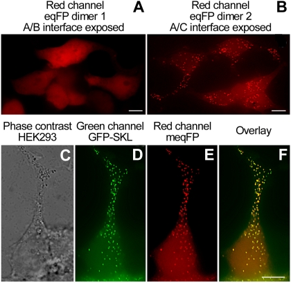
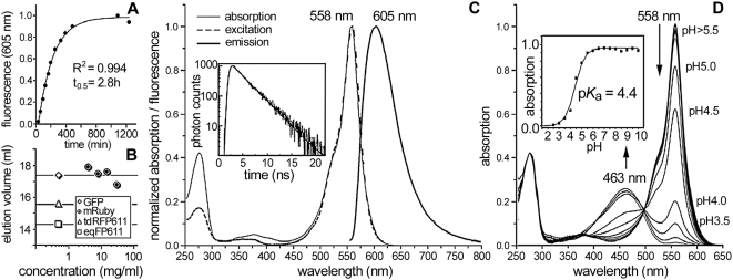
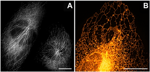
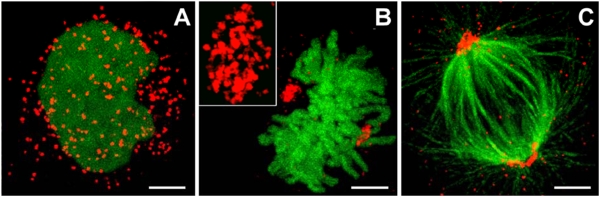
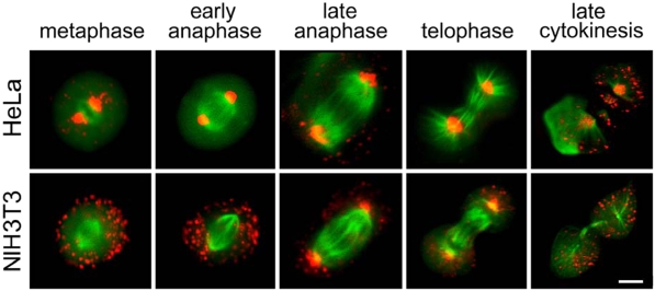
A monomeric variant of the red fluorescent protein eqFP611, mRuby, is described. With excitation and emission maxima at 558 nm and 605 nm, respectively, and a large Stokes shift of 47 nm, mRuby appears particularly useful for imaging applications. The protein shows an exceptional resistance to denaturation at pH extremes. Moreover, mRuby is about ten-fold brighter compared to EGFP when being targeted to the endoplasmic reticulum. The engineering process of eqFP611 revealed that the C-terminal tail of the protein acts as a natural peroxisomal targeting signal (PTS). Using an mRuby variant carrying the eqFP611-PTS, we discovered that ordered inheritance of peroxisomes is widespread during mitosis of different mammalian cell types. The ordered partitioning is realized by the formation of peroxisome clusters around the poles of the mitotic spindle and ensures that equal numbers of the organelle are inherited by the daughter cells. The unique spectral properties make mRuby the marker of choice for a multitude of cell biological applications. Moreover, the use of mRuby has allowed novel insights in the biology of organelles responsible for severe human diseases.


Conflict of interest statement

Competing Interests: A US patent application covering mRuby and its applications is pending.

Figures

Figure 1
Figure 1. Subcellular localization of eqFP611 variants in HEK293 cells.
(A) The eqFP dimer 1 (RFP611 T122R) is evenly distributed over the cell. (B) The enhanced eqFP dimer 2 (T122G, F194A) shows a granular localization. (C–F) Cells co-expressing EGFP-SKL and an enhanced monomeric eqFP611 (T122R, F194A) variant show co-localization of green and red fluorescence in the peroxisomes. (C) Phase contrast image. (D) Green-channel image acquired with a standard FITC filter set. (E) Red-channel image produced with a standard TRITC filter set. (F) Overlay image of the green and red channels. Bars: 2.5 µm.
Figure 2
Figure 2. Characterization of mRuby properties.
(A) In vitro chromophore maturation at 37°C as determined from the fluorescence emission at 605 nm (solid line: exponential fit). (B) Elution volume for concentrations of 4–30 mg/ml (0.16–1.2 mM) during gel-filtration analysis (standards for the oligomerization degree: GFP S65T, monomer; td-RFP611, dimer; eqFP611, tetramer). (C) Absorption, excitation and emission spectra (inset: fluorescence decay with a single exponential fit, yielding a lifetime of 2.6±0.1 ns). (D) Absorption spectra at various pH values (inset: absorption at 558 nm versus pH; Henderson-Hasselbalch fit reveals a pKa of 4.4).
Figure 3
Figure 3. Applications of mRuby as a cellular marker protein.
(A) Epifluorescence image of NIH3T3 cells expressing mRuby fused to human α-tubulin. Two cells from a single image were arranged in closer proximity (B) Spinning-disk confocal image of the endoplasmic reticulum of a HeLa cell stained with ER-mRuby-KDEL. False colors encode fluorescence intensity. Bars: 2.5 µm.
Figure 4
Figure 4. Peroxisome clusters in HEK293 cells.
(A–C) Confocal images (Leica TCS 4Pi) of HEK293 cells co-expressing mRuby-PTS and H2B-EGFP during (A) interphase and (B) metaphase. The inset in (B) shows a magnification of a peroxisomal cluster. Tubulin fibers are labelled in green with Alexa Fluor©-488 via an anti-α-tubulin antibody (C) to reveal peroxisomal clustering at the spindle poles. Bars: 2 µm.
Figure 5
Figure 5. Inheritance of peroxisomes.
Distribution of peroxisomes during different stages of mitosis in HeLa (upper row) and NIH3T3 cells (lower row). Peroxisomes are highlighted by mRuby-PTS. The spindle of the mitotic cells is stained with EGFP fused to a tubulin-binding protein . Images were taken 24–48 h after transfection on an Olympus IX71 microscope equipped with standard FITC and TRITC filter sets. Bar: 5 µm.

References

    1. Shaner NC, Patterson GH, Davidson MW. Advances in fluorescent protein technology. J Cell Sci. 2007;120:4247–4260. - PubMed
    1. Matz MV, Fradkov AF, Labas YA, Savitsky AP, Zaraisky AG, et al. Fluorescent proteins from nonbioluminescent Anthozoa species. Nat Biotechnol. 1999;17:969–973. - PubMed
    1. Wiedenmann J. The application of an orange fluorescent protein and further colored proteins and the corresponding genes from the species group Anemonia sp. (sulcata) Pennant, (Cnidaria, Anthozoa, Actinaria) in gene technology and molecular biology. (Die Anwendung eines orange fluoreszierenden Proteins und weiterer farbiger Proteine und der zugehörenden Gene aus der Artengruppe Anemonia sp. (sulcata) Pennant, (Cnidaria, Anthozoa, Actinaria) in Gentechnologie und Molekularbiologie.). Patent DE 197 18 640 Deutsches Patent- und Markenamt. Deutschland. 1997:1–18.
    1. Wiedenmann J, Schenk A, Rocker C, Girod A, Spindler KD, et al. A far-red fluorescent protein with fast maturation and reduced oligomerization tendency from Entacmaea quadricolor (Anthozoa, Actinaria). Proc Natl Acad Sci U S A. 2002;99:11646–11651. - PMC - PubMed
    1. Merzlyak EM, Goedhart J, Shcherbo D, Bulina ME, Shcheglov AS, et al. Bright monomeric red fluorescent protein with an extended fluorescence lifetime. Nat Methods. 2007;4:555–557. - PubMed

Publication types

Substances