Clinical Trial
. 2009 Jan;65(1):98-107.
doi: 10.1002/ana.21526.

Taurine deficiency is a cause of vigabatrin-induced retinal phototoxicity

Affiliations
Clinical Trial

Taurine deficiency is a cause of vigabatrin-induced retinal phototoxicity

Firas Jammoul et al. Ann Neurol. 2009 Jan.

Abstract

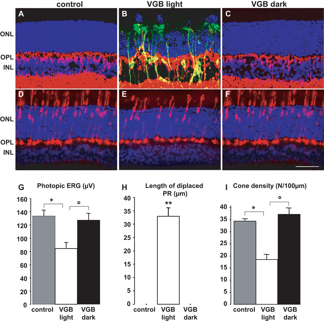
Objective: Although vigabatrin irreversibly constricts the visual field, it remains a potent therapy for infantile spasms and a third-line drug for refractory epilepsies. In albino animals, this drug induces a reduction in retinal cell function, retinal disorganization, and cone photoreceptor damage. The objective of this study was to investigate the light dependence of the vigabatrin-elicited retinal toxicity and to screen for molecules preventing this secondary effect of vigabatrin.

Methods: Rats and mice were treated daily with 40 and 3mg vigabatrin, respectively. Retinal cell lesions were demonstrated by assessing cell function with electroretinogram measurements, and quantifying retinal disorganization, gliosis, and cone cell densities.

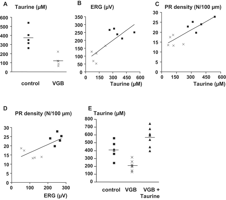
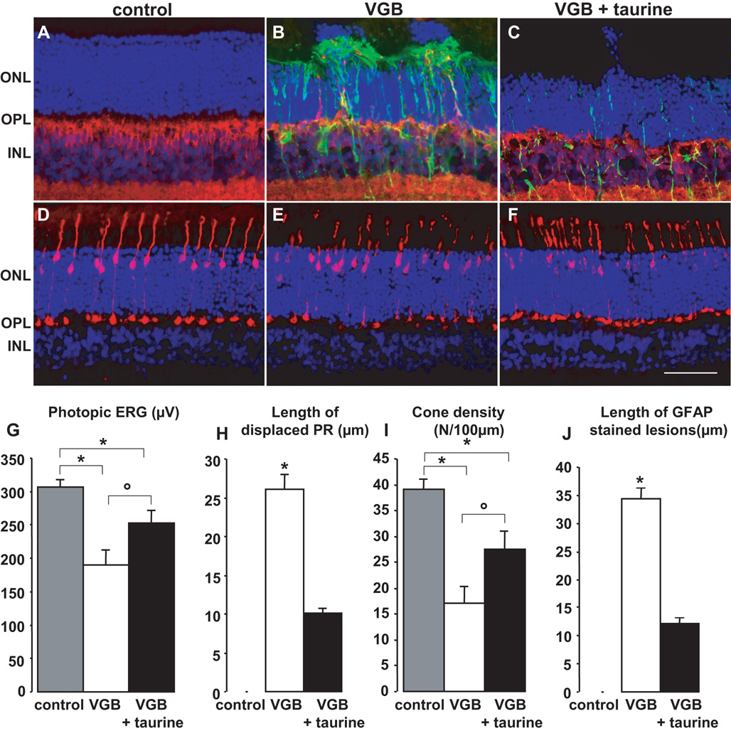
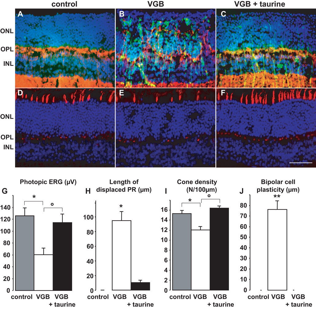
Results: Vigabatrin-elicited retinal lesions were prevented by maintaining animals in darkness during treatment. Different mechanisms including taurine deficiency were reported to produce such phototoxicity; we therefore measured amino acid plasma levels in vigabatrin-treated animals. Taurine levels were 67% lower in vigabatrin-treated animals than in control animals. Taurine supplementation reduced all components of retinal lesions in both rats and mice. Among six vigabatrin-treated infants, the taurine plasma level was found to be below normal in three patients and undetectable in two patients.

Interpretation: These results indicate that vigabatrin generates a taurine deficiency responsible for its retinal phototoxicity. Future studies will investigate whether cotreatment with taurine and vigabatrin can limit epileptic seizures without inducing the constriction of the visual field. Patients taking vigabatrin could gain immediate benefit from reduced light exposures and dietetic advice on taurine-rich foods.


Conflict of interest statement

Conflicting interests: Patents were filed on the present results by INSERM U-592 (Institut de la Vision). INSERM U592 received financial support from the company Ovation Pharmaceuticals for the present work. Stephen Collins is an employee of Ovation pharmaceuticals, which has applied to the American Food and Drug Administration (FDA) for the use of vigabatrin in the USA for infantile spasms and refractory complex partial seizures in adults.

Figures

Figure 1
Figure 1. The in vivo retinal phototoxicity of vigabatrin
(A–F) Retinal sections showing the lesions in a rat treated with VGB for 45 days in room light (B, E, VGB light) and absence of lesions in a VGB-treated rat maintained in darkness (C, F, VGB dark) and in a control animal (A, D). Sections were stained with the nuclear dye, DAPI, (blue in A–F) and immunolabelled for GFAP (green in A–C), Goα (red in A–C) and cone arrestin (red in D–F). Photoreceptor nuclei displaced above the outer nuclear layer (ONL), Goα-positive bipolar cell dendrites sprouting into the ONL and GFAP-positive processes extending vertically throughout the retina are only observed in rats treated with VGB in the 12h/12h light/dark cycle (B), not in control animals (A) or VGB-treated animals maintained in darkness (C). Similarly, fewer cone arrestin-positive photoreceptors and their inner/outer segments were observed in areas with normal retinal layering in VGB-treated rats exposed to a 12h/12h light/dark cycle (E), than were observed for control (D) or VGB-treated rats maintained in the dark (F). Quantification of photopic ERG amplitudes (G), lengths of retinal areas with displaced photoreceptor (PR) nuclei (H) and cone inner/outer segment density (I) in control rats (s.e.m., n=9), for VGB-treated animals either exposed to a 12h/12h light/dark cycle (VGB light, s.e.m., n= 10) or maintained in darkness (VGB dark, s.e.m., n= 10). The scale bar represents 50µm (IPL: inner plexiform layer).
Figure 2
Figure 2. Correlation between taurine levels and the phototoxicity of vigabatrin
A) Taurine plasma levels in control and treated animals in each group. B) Correlation between taurine plasma levels and the photopic ERG amplitude in VGB-treated control rats. The correlation factor between these two factors is equal to 0.769 (p=0.0093%, n=10). C) Correlation between the taurine plasma levels and the density of inner/outer cone photoreceptor segments. The correlation factor between these two parameters is equal to 0.818 (p=0.0038%, n=10). D) Correlation between the photopic ERG amplitudes and the density of inner/outer cone photoreceptor segments. The correlation factor between these two factors is equal to 0.703 (p=0.0023%, n=10). E) Recovery of taurine plasma levels by taurine supplementation in VGB-treated rats. In A and E, the horizontal line represents the mean value.
Figure 3
Figure 3. Partial prevention of vigabatrin-induced retinal toxicity in rats by taurine supplementation
(A–F) Retinal sections showing that VGB-elicited retinal lesions are less extensive in a rat with taurine supplementation (C, F, VGB + taurine) than without (B, E, VGB), but still greater than in a control animal (A, D). These sections were stained with DAPI (blue in A–F) and immunolabelled with antibodies directed against Goα (red in A–C), GFAP (green in A–C) and cone arrestin (red in D–F). Photoreceptor nuclei displaced above the outer nuclear layer (ONL) are observed in both groups of VGB-treated rats treated with or without taurine supplementation (B, C), but not in control animals (A). GFAP-positive processes extending vertically throughout the retina were observed in VGB-treated rats (B), but not in control animals (A); higher levels of GFAP staining were also observed in VGB-treated rats receiving taurine supplementation (C) than those observed in control animals (A). Similarly, there were clearly fewer cone arrestin-positive photoreceptors in the VGB-treated rats receiving morning injections (E) than in control animals (D), with a smaller decrease in cone arrestin-positive photoreceptors in VGB-treated rats receiving evening injections (F). Quantification of photopic ERG amplitude (G), length of retinal areas with displaced photoreceptor (PR) nuclei (H), density of cone inner/outer segments (I) and areas with increased GFAP expression (J) in control rats (s.e.m., n=6), in the VGB-treated animals with or without taurine supplementation (VGB, n=7; VGB + taurine, n=7, s.e.m.). The scale bar represents 50µm (IPL: inner plexiform layer).
Figure 4
Figure 4. Partial prevention of vigabatrin-retinal toxicity in mice by taurine suppementation
(A–F) Retinal sections showing that VGB-elicited retinal lesions are less extensive in a mouse with a taurine supplementation (C, F, VGB + taurine) than without (B, E, VGB), but still greater than in a control animal (A, D). These sections were immunolabelled with antibodies directed against Goα (red in A–C) and GFAP (green in A–C) and stained with a peanut lectin (PNA, red in D–F) and DAPI (blue in A–F). Photoreceptor nuclei displaced above the outer nuclear layer (ONL) were only observed in the VGB-treated mice (B), not in control animals (A) or VGB-treated mice receiving taurine supplementation. GFAP-positive processes extending vertically throughout the retina were observed in the VGB-treated animals (B), and increased GFAP staining was also observed in VGB-treated mice with taurine supplementation (C), compared to control animals (A). Similarly, there were fewer PNA-positive inner/outer segments of photoreceptors in VGB-treated mice (E) than in either control animals (D) or VGB-treated mice with taurine supplementation. Quantification of photopic ERG amplitude (G), length of retinal areas with displaced photoreceptor (PR) nuclei (H), density of cone inner/outer segments (I) and areas with bipolar cell sprouting into the ONL (J) in control mice (s.e.m., n=5), in VGB-treated animals with or without taurine supplementation (VGB, s.e.m., n= 7; VGB + taurine, s.e.m., n= 7). The scale bar represents 50µm (IPL: inner plexiform layer).

References

    1. Ben-Menachem E, Dulac O, Chiron C. Vigabatrin. In: Engel Jerome, Jr, Pedley Timothy A., editors. Epilepsy: a comprehensive text book. Second edition. Philadelphia: Lippincott Williams & Wilkins; 2008. pp. 1683–1693.
    1. Krauss GL, Johnson MA, Miller NR. Vigabatrin-associated retinal cone system dysfunction: electroretinogram and ophthalmologic findings. Neurology. 1998;50:614–618. - PubMed
    1. Ruether K, Pung T, Kellner U, et al. Electrophysiologic evaluation of a patient with peripheral visual field contraction associated with vigabatrin. Archives of ophthalmology. 1998;116:817–819. - PubMed
    1. Eke T, Talbot JF, Lawden MC. Severe persistent visual field constriction associated with vigabatrin. BMJ (Clinical research ed. 1997;314:180–181. - PMC - PubMed
    1. Johnson MA, Krauss GL, Miller NR, et al. Visual function loss from vigabatrin: effect of stopping the drug. Neurology. 2000;55:40–45. - PubMed

Publication types

MeSH terms