

Solute transport in cyclically deformed porous tissue scaffolds with controlled pore cross-sectional geometries
- PMID: 19196145
- PMCID: PMC2792109
- DOI: 10.1089/ten.tea.2008.0382
Solute transport in cyclically deformed porous tissue scaffolds with controlled pore cross-sectional geometries
Abstract
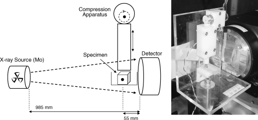
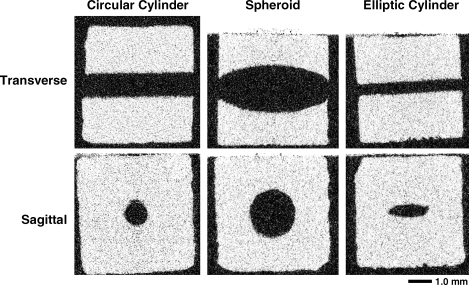
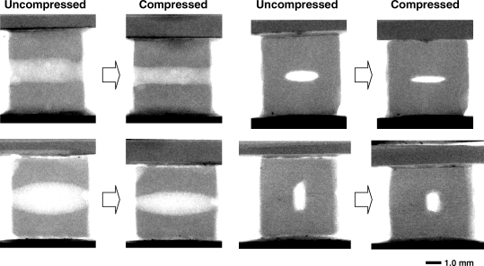
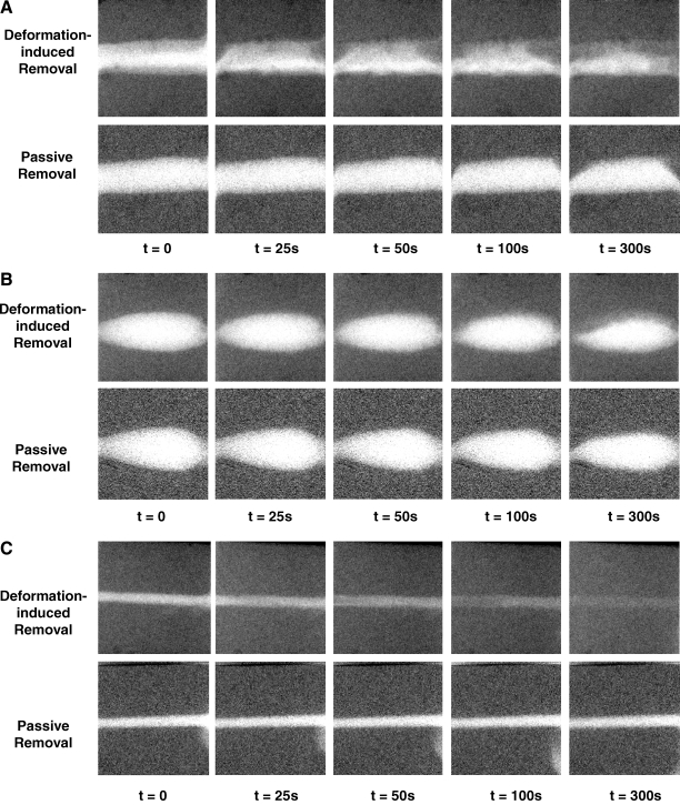
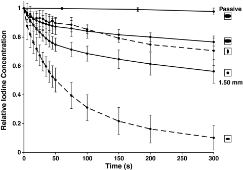
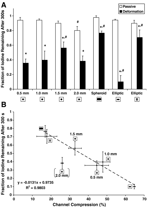
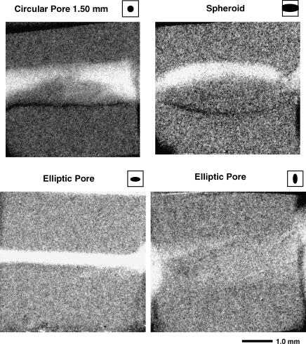
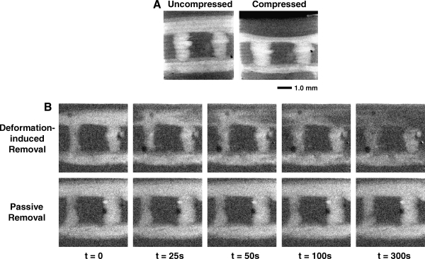
The objective of this study was to investigate the influence of pore geometry on the transport rate and depth after repetitive mechanical deformation of porous scaffolds for tissue engineering applications. Flexible cubic imaging phantoms with pores in the shape of a circular cylinder, elliptic cylinder, and spheroid were fabricated from a biodegradable polymer blend using a combined 3D printing and injection molding technique. The specimens were immersed in fluid and loaded with a solution of a radiopaque solute. The solute distribution was quantified by recording 20 microm pixel-resolution images in an X-ray microimaging scanner at selected time points after intervals of dynamic straining with a mean strain of 8.6+/-1.6% at 1.0 Hz. The results show that application of cyclic strain significantly increases the rate and depth of solute transport, as compared to diffusive transport alone, for all pore shapes. In addition, pore shape, pore size, and the orientation of the pore cross-sectional asymmetry with respect to the direction of strain greatly influence solute transport. Thus, pore geometry can be tailored to increase transport rates and depths in cyclically deformed scaffolds, which is of utmost importance when thick, metabolically functional tissues are to be engineered.
Figures
Similar articles
-
Validation of a fluid-structure interaction model of solute transport in pores of cyclically deformed tissue scaffolds.Tissue Eng Part C Methods. 2010 Oct;16(5):1145-56. doi: 10.1089/ten.TEC.2009.0685. Tissue Eng Part C Methods. 2010. PMID: 20136371 Free PMC article.
-
Cyclic deformation-induced solute transport in tissue scaffolds with computer designed, interconnected, pore networks: experiments and simulations.Ann Biomed Eng. 2009 Aug;37(8):1601-12. doi: 10.1007/s10439-009-9712-3. Epub 2009 May 23. Ann Biomed Eng. 2009. PMID: 19466547 Free PMC article.
-
Permeability and mechanical properties of gradient porous PDMS scaffolds fabricated by 3D-printed sacrificial templates designed with minimal surfaces.Acta Biomater. 2019 Sep 15;96:149-160. doi: 10.1016/j.actbio.2019.06.040. Epub 2019 Jun 25. Acta Biomater. 2019. PMID: 31252172
-
Quasi-static and dynamic in vitro mechanical response of 3D printed scaffolds with tailored pore size and architectures.Mater Sci Eng C Mater Biol Appl. 2019 Mar;96:176-182. doi: 10.1016/j.msec.2018.11.019. Epub 2018 Nov 15. Mater Sci Eng C Mater Biol Appl. 2019. PMID: 30606523
-
The first systematic analysis of 3D rapid prototyped poly(ε-caprolactone) scaffolds manufactured through BioCell printing: the effect of pore size and geometry on compressive mechanical behaviour and in vitro hMSC viability.Biofabrication. 2013 Dec;5(4):045004. doi: 10.1088/1758-5082/5/4/045004. Epub 2013 Nov 6. Biofabrication. 2013. PMID: 24192056
Cited by
-
Validation of a fluid-structure interaction model of solute transport in pores of cyclically deformed tissue scaffolds.Tissue Eng Part C Methods. 2010 Oct;16(5):1145-56. doi: 10.1089/ten.TEC.2009.0685. Tissue Eng Part C Methods. 2010. PMID: 20136371 Free PMC article.
-
An in vitro model for the pathological degradation of articular cartilage in osteoarthritis.J Biomech. 2014 Feb 7;47(3):645-52. doi: 10.1016/j.jbiomech.2013.11.050. Epub 2013 Dec 10. J Biomech. 2014. PMID: 24360770 Free PMC article.
-
Long-term in vivo survival of 3D-bioprinted human lipoaspirate-derived adipose tissue: proteomic signature and cellular content.Adipocyte. 2022 Dec;11(1):34-46. doi: 10.1080/21623945.2021.2014179. Adipocyte. 2022. PMID: 34957918 Free PMC article.
References
-
- Mygind T. Stiehler M. Baatrup A. Li H. Zou X. Flyvbjerg A. Kassem M. Bunger C. Mesenchymal stem cell ingrowth and differentiation on coralline hydroxyapatite scaffolds. Biomaterials. 2007;28:1036. - PubMed
-
- Carrier R.L. Rupnick M. Langer R. Schoen F.J. Freed L.E. Vunjak-Novakovic G. Perfusion improves tissue architecture of engineered cardiac muscle. Tissue Eng. 2002;8:175. - PubMed
-
- Karande T.S. Ong J.L. Agrawal C.M. Diffusion in musculoskeletal tissue engineering scaffolds: design issues related to porosity, permeability, architecture, and nutrient mixing. Ann Biomed Eng. 2004;32:1728. - PubMed
-
- Ramrattan N.N. Heijkants R.G. van Tienen T.G. Schouten A.J. Veth R.P. Buma P. Assessment of tissue ingrowth rates in polyurethane scaffolds for tissue engineering. Tissue Eng. 2005;11:1212. - PubMed
-
- Silva M.M. Cyster L.A. Barry J.J. Yang X.B. Oreffo R.O. Grant D.M. Scotchford C.A. Howdle S.M. Shakesheff K.M. Rose F.R. The effect of anisotropic architecture on cell and tissue infiltration into tissue engineering scaffolds. Biomaterials. 2006;27:5909. - PubMed
Publication types
MeSH terms
Substances
Grants and funding
LinkOut - more resources
Full Text Sources
Other Literature Sources
