. 2009 May 1;419(3):595-602.
doi: 10.1042/BJ20081449.

The C-terminus of the P22 tailspike protein acts as an independent oligomerization domain for monomeric proteins

Affiliations

The C-terminus of the P22 tailspike protein acts as an independent oligomerization domain for monomeric proteins

Tawnya Webber et al. Biochem J. .

Abstract

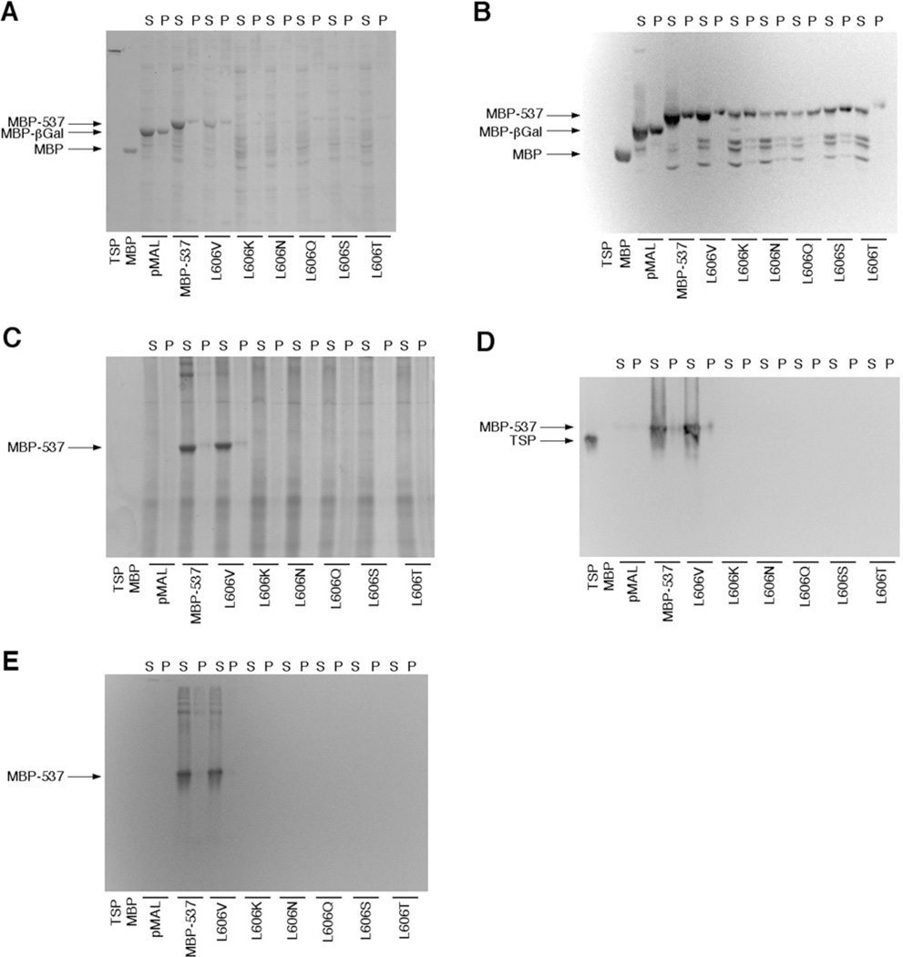
TSP (P22 tailspike protein) is a well-established model system for studying the folding and assembly of oligomeric proteins, and previous studies have documented both in vivo and in vitro folding intermediates using this protein. Especially important is the C-terminus of TSP, which plays a critical role in the assembly and maturation of the protrimer intermediate to its final trimeric form. In the present study, we show that by grafting the C-terminus of TSP on to the monomeric MBP (maltose-binding protein), the resulting chimaera (MBP-537) is a trimeric protein. Moreover, Western blot studies (using an anti-TSP antibody) indicate that the TSP C-terminus in the MBP-537 chimaera has the same conformation as the native TSP. The oligomerization of the MBP-537 chimaera appears to involve hydrophobic interactions and a refolding sequence, both of which are analogous to the native TSP. These results underscore the importance of the TSP C-terminus in the assembly of the mature trimer and demonstrate its potential utility as a model to study the folding and assembly of the TSP C-terminus in isolation.


Figures

Figure 1
Figure 1. Structure and folding pathway of TSP
(A) Ribbon diagram of the structure of TSP. The four main structural features are indicated on the structure. (B) Ribbon diagram of the C-terminus, orientated to view the structure down the long axis of the protein from the β-helix domain to the end of the protein. Sheets D and E are indicated with arrows. (C) In vitro folding and aggregation pathway of the TSP. Unfolded monomer folds and forms either aggregate-prone monomer (bottom) or folding competent monomer (top). Adapted from [3] with permission © (2003) Wiley.
Figure 2
Figure 2. Comparison of the electrophoretic mobility of MBP-537 chimaera compared to native TSP
Purified protein (0.5 µg/ml) was separated by SDS/PAGE (A) or non-denaturing PAGE (B) and stained with Coomassie Blue. Lane 1, TSP; lane 2, purified MBP-537; lane 3, MBP-537 digested overnight with Genenase™ I. Lane 4 in (A) is the molecular mass markers, with values in kDa to the right. All lanes were from the same gels and were grouped together for publication purposes. Dividing lines indicate where each gel was merged.
Figure 3
Figure 3. Sedimentation velocity and sedimentation equilibrium analysis of the MBP-537 chimaera
(A) Raw sedimentation velocity data (grey line) fitted to a monodisperse species (black line). The calculated (SEDFIT) sedimentation coefficient is 7.25, which is consistent with a species having a molecular mass of 165.7 kDa. (B) Equilibrium sedimentation data (circles, 10 000 rev./min; diamonds, 8000 rev./min) were analysed with SEDPHAT using a model for monodisperse protein, which gives a calculated species molecular mass of 170 kDa. The fit of the data is shown as the lines through the points, with residuals plotted below the graphs.
Figure 4
Figure 4. Comparison of antibody reactivity of the chimaeric MBP-537 with antibody reactivity of the TSP and monomeric MBP
Purified protein (0.1 µg/ml) was separated by SDS/PAGE (A) or non-denaturing PAGE (B) and transferred on to nitrocellulose for Western blotting. Lane 1, TSP; lane 2, purified MBP-537; lane 3, MBP-537 chimaera that has been digested overnight with Genenase™ I to separate the MBP from the TSP C-terminus. The blots on the left were incubated with anti-MBP antibody (NEB). The blots on the right were incubated with the mAB155 (anti-TSP) antibody. All lanes were from the same blots and were grouped together for publication purposes. Dividing lines indicate where each blot was merged.
Figure 5
Figure 5. SDS/PAGE and Western blot analysis of mutated MBP-537 chimaeras
Whole-cell lysates from expressions of MBP-537 chimaeras containing various mutations at Leu606 (TSP numbering) were analysed using both SDS/PAGE and native PAGE. (A) Coomassie Blue-stained SDS/PAGE gel of whole-cell lysates. (B) Western blot of SDS/PAGE-separated lysates treated with an anti-MBP antibody. (C) Coomassie Blue-stained native gel of whole-cell lysates. (D) Western blot of native PAGE-separated lysates treated with an anti-TSP antibody. (E) Western blot of native PAGE-separated lysates treated with an anti-MBP antibody. Lane 1 of each gel contains purified TSP and lane 2 contains purified monomeric MBP. The remaining lanes contain the soluble portion (S) and pellet (P) from each lysate. Lanes 3 and 4 contain the soluble and pellet fractions from an expression of the empty pMal plasmid, which expresses a MBP–βGal fusion protein. Lanes 5 and 6 contain the soluble and pellet fractions from expression of the native MBP-537 chimaera. The soluble and pellet fractions from expressions of individual mutant MBP-537 chimaeras were separated in lanes 7–18, with the individual mutation indicated over each pair of lanes.
Figure 6
Figure 6. Analysis of MBP-537 refolding
(A) Denatured MBP-537 and TSP were refolded at 100 µg/ml and 20°C. Samples were collected, quenched on ice and analysed by non-denaturing PAGE. The time each sample was collected is shown between the two gels. The various refolding species are indicated on the right side of each gel. A similar progression from monomer to dimer to the final form is seen with both proteins. Interestingly, an early intermediate similar to the TSP protrimer is seen in the MBP-537 sample. (B) TSP and MBP-537 protein were refolded at 100 µg/ml and 0°C for 4 h. The samples were separated on a native gel and the gel was silver-stained. Lane 1, refolded TSP; lane 2, refolded MBP-537; lane 3, blank; lane 4, purified MBP-537; lane 5, purified TSP. TSP primarily forms a dimer and protrimer, whereas the MBP-537 chimaera forms primarily monomer. TSP dimer and mature trimer migrate very similarly on 7 % native gels due to similar Stokes radii [25].

References

    1. Steinbacher S, Seckler R, Miller S, Steipe B, Huber R, Reinemer P. Crystal structure of P22 tailspike protein: interdigitated subunits in a thermostable trimer. Science. 1994;265:383–385. - PubMed
    1. Miller S, Schuler B, Seckler R. Phage P22 tailspike protein: removal of head-binding domain unmasks effects of folding mutations of native thermal stability. Protein Sci. 1998;7:2223–2232. - PMC - PubMed
    1. Gage MJ, Robinson AS. C-terminal hydrophobic interactions play a critical role in oligomeric assembly of the P22 tailspike trimer. Protein Sci. 2003;12:2732–2747. - PMC - PubMed
    1. Seckler R, Fuchs A, King J, Jaenicke R. Reconstitution of the thermostable trimeric phage P22 tailspike from denatured chains in vitro. J. Biol. Chem. 1989;264:11750–11753. - PubMed
    1. Miller S, Schuler B, Seckler R. A reversibly unfolding fragment of P22 tailspike protein with native structure: the isolated β-helix domain. Biochemistry. 1998;37:9160–9168. - PubMed

Publication types

MeSH terms