. 2009 Mar;19(3):415-26.
doi: 10.1101/gr.084509.108. Epub 2009 Feb 4.

Into the blue: gene duplication and loss underlie color vision adaptations in a deep-sea chimaera, the elephant shark Callorhinchus milii

Affiliations

Into the blue: gene duplication and loss underlie color vision adaptations in a deep-sea chimaera, the elephant shark Callorhinchus milii

Wayne L Davies et al. Genome Res. 2009 Mar.

Abstract

The cartilaginous fishes reside at the base of the gnathostome lineage as the oldest extant group of jawed vertebrates. Recently, the genome of the elephant shark, Callorhinchus milii, a chimaerid holocephalan, has been sequenced and therefore becomes the first cartilaginous fish to be analyzed in this way. The chimaeras have been largely neglected and very little is known about the visual systems of these fishes. By searching the elephant shark genome, we have identified gene fragments encoding a rod visual pigment, Rh1, and three cone visual pigments, the middle wavelength-sensitive or Rh2 pigment, and two isoforms of the long wavelength-sensitive or LWS pigment, LWS1 and LWS2, but no evidence for the two short wavelength-sensitive cone classes, SWS1 and SWS2. Expression of these genes in the retina was confirmed by RT-PCR. Full-length coding sequences were used for in vitro expression and gave the following peak absorbances: Rh1 496 nm, Rh2 442 nm, LWS1 499 nm, and LWS2 548 nm. Unusually, therefore, for a deep-sea fish, the elephant shark possesses cone pigments and the potential for trichromacy. Compared with other vertebrates, the elephant shark Rh2 and LWS1 pigments are the shortest wavelength-shifted pigments of their respective classes known to date. The mechanisms for this are discussed and we provide experimental evidence that the elephant shark LWS1 pigment uses a novel tuning mechanism to achieve the short wavelength shift to 499 nm, which inactivates the chloride-binding site. Our findings have important implications for the present knowledge of color vision evolution in early vertebrates.


Figures

Figure 1.
Figure 1.
Phylogenetic analysis of LWS1, LWS2, Rh2, and Rh1 elephant shark vertebrate retinal opsin sequences and coding sequences of other related vertebrate species. (A) A neighbor-joining tree was constructed (Saitou and Nei 1987) by applying a Kimura 2-parameter substitution matrix (Kimura 1980) with 1000 bootstrapping replications (degree of support for internal branching is shown as a percentage at the base of each node). (B) A maxiumum likelihood tree was generated with a Kimura 2-parameter substitution matrix (Kimura 1980), a gamma distribution parameter of 1 using PHYML (Guindon et al. 2005) and bootstrapping with 500 replicates (degree of support for internal branching is shown as a percentage at the base of each node). (C) A Bayesian probabilistic inference method was performed with a Metropolis Markov chain Monte Carlo algorithm (Huelsenbeck and Ronquist 2001; Ronquist and Huelsenbeck 2003). A general time-reversal model (Lanave et al. 1984) was used with posterior probability values (represented as a percentage) indicated at the base of each node. The scale bar indicates the number of nucleotide substitutions per site. The Drosophila melanogaster (fruit fly) Rh4 (accession no. NM057353) was used as an outgroup. The sequences used for generating the tree are as follows: (a) RhA/Rh1 opsin class: zebrafish (Danio rerio), NM131084; goldfish (Carassius auratus), L11863; guppy (Poecilia reticulata), Y11147; John Dory (Zeus faber), Y14484; smaller spotted catshark (Scyliorhinus canicula), Y17585; dogfish (Galeus melastomus), Y17586; little skate (Raja erinacea), U81514; elephant shark (Callorhinchus milii), EF565167; sea lamprey (Petromyzon marinus) (RhA), AH005459; Arctic lamprey (Lethenteron japonica) (RhA), M63632; pouched lamprey (Geotria australis) (RhA), AY366493; (b) RhB/Rh2 opsin class: zebrafish (Danio rerio), AB087805 (Rh2.1), AB087806 (Rh2.2), AB087807 (Rh2.3), AB087808 (Rh2.4); goldfish (Carassius auratus), L11865 (Rh2.1), L11866 (Rh2.2); bluefin killifish (Lucania goodei), AY296739; rainbow trout (Oncorhynchus mykiss), AF425072; elephant shark (Callorhinchus milii), EF565168; pouched lamprey (Geotria australis) (RhB), AY366494; (c) SWS2 opsin class: zebrafish (Danio rerio), NM131192; goldfish (Carassius auratus), L11864; bluefin killifish (Lucania goodei), AY296737; rainbow trout (Oncorhynchus mykiss), AF425075; pouched lamprey (Geotria australis), AY366492; (d) SWS1 opsin class: zebrafish (Danio rerio), NM131319; goldfish (Carassius auratus), D85863; pouched lamprey (Geotria australis), AY366495; (e) LWS opsin class: zebrafish (Danio rerio), NM131175; goldfish (Carassius auratus), L11867; pouched lamprey (Geotria australis), AY366491; elephant shark (Callorhinchus milii), EF565165 (LWS1), EF565166 (LWS2).
Figure 2.
Figure 2.
Absorption spectra of regenerated C. milii (A) rod (Rh1) and (B) cone (Rh2, LWS1, and LWS2) visual pigments. For Rh1 pigments, representative dark (●) and bleached spectra (○) are shown, with difference spectra that have been fitted with a Govardovskii et al. (2000) template (line) in the inset to determine the λmax value. For Rh2, LWS1, and LWS2 pigments, representative difference spectra are shown that were fitted to Govardovskii et al. (2000) templates to determine the λmax values. For LWS1 pigments, the spectral peak of absorbance was determined in the presence (continuous line) and absence of chloride ions (dotted line).
Figure 3.
Figure 3.
Alignment of the amino acid sequences of visual pigments expressed in the elephant shark (Callorhinchus milii; CM) and the common cow (Bos taurus, BT). (Rh1) Rod opsin; (LWS1, LWS2) long-wavelength-sensitive 1 and 2; (Rh2) cone rhodopsin 2 (middle-wavelength-sensitive). (*) An identical consensus residue between all four elephant shark opsin sequences and bovine Rh1. (: and .) A conserved or semiconservative amino substitution, respectively, with the codon-matched protein alignment. Gaps inserted to maintain a high degree of identity present between the opsin sequences derived from C. milii retina and bovine Rh1 are indicated by dashes (–). Seven putative transmembrane domains (TMDs) are indicated by gray shading. The TMDs shown for bovine rhodopsin were determined by crystallography (Palczewski et al. 2000). The putative positions of the TMDs for each elephant shark visual pigment were predicted online using TMHMM Server Version 2.0 (http://www.cbs.dtu.dk/services/TMHMM/). Residues identified as being critical for correct opsin protein conformation (boxed). Comparison of the opsin amino acid sequences of C. milii and bovine rod opsin demonstrated that the critical residues involved in the maintenance of the tertiary structure of the opsin molecule are present. With the use of the conventional numbering system of the bovine rod opsin polypetide sequence, these key sites include (1) three conserved cysteine (C) residues at positions 110 (TMD3), 185 (ECD2), and 187 (ECD2) that are involved in disulphide bond formation (Karnik and Khorana 1990), except for a Thr (T) residue at position 185 in the elephant shark LWS1 and LWS2 opsins, which are also conserved throughout the rest of the vertebrate LWS opsin class; (2) a conserved glutamate (E) at position 113 (TMD3) that provides the negative counterion to the proton of the Schiff base (Sakmar et al. 1989); (3) a conserved glutamate (E) at position 134 (TM3) that provides a negative charge to stabilize the inactive opsin molecule (Cohen et al. 1993); (4) a conserved lysine (K) at position 296 (TM7) that is covalently linked to the chromophore via a Schiff base (Dratz and Hargrave 1983); (5) conservation of two cysteine (C) residues at putative palmitoylation positions 322 and 323 (Ovchinnikov et al. 1988) in both elephant shark Rh1 and Rh2 opsins, but not LWS1 and LWS2 opsins; (6) the presence of a number of Ser (S) and Thr (T) residues in the carboxyl terminus, which are potential targets for phosphorylation by rhodopsin kinases in the deactivation of metarhodopsin II (Palczewski et al. 1993; Sakmar and Fahmy 1996; Zhao et al. 1997); and (7) the conserved glycosylation sites at positions 2 and 15 (Sakmar and Fahmy 1996) in the MWS opsins (Rh1 and Rh2) identified in the retina of C. milii.
Figure 4.
Figure 4.
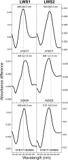
Absorption spectra of regenerated C. milii LWS1 (left) and LWS2 (right) mutants. (Top) His181Tyr single substitutions in both LWS1 and LWS2 pigments. (Middle) Ser292Ala (LWS1) or Ala292Ser (LWS2) single substitutions. (Bottom) His181Tyr and Ser292Ala (LWS1) or His181Tyr and Ala292Ser (LWS2) double substitutions. For all pigments, representative difference spectra are shown that were fitted to Govardovskii et al. (2000) templates to determine the λmax values. The vertical dotted line shows the relative position of the spectral peak values for both wild-type LWS1 (499 nm) and LWS2 (548 nm) regenerated pigments.
Figure 5.
Figure 5.
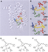
(A) Structural model of elephant shark LWS visual pigments showing the relative position of the five key LWS tuning sites (164, 181, 261, 269, and 292) within the retinal-binding pocket containing the retinylidene chromophore (yellow), the Lys296 chromophore attachment site via a Schiff base (blue), and the Glu113 counterion (orange). LWS tuning sites are color coded whether they are situated proximally to (green) or distally (red) from the Schiff base. Enlarged view showing the key residues of (B) LWS1 and (C) LWS2 pigments surrounding the retinylidene chromophore. The model was created using Swiss Model (Guex and Peitsch 1997) and is based on the crystal structure of bovine rhodopsin (Palczewski et al. 2000). A schematic representation of the proposed model for the interaction between residues 181 and 292 in close proximity to the Schiff base of the chromophore, showing (D) a chloride ion acting as a counterion to the positive charge present on His181, (E) the loss of binding and repulsion of a chloride ion due to the net negative polar charge of the Tyr residue at position 181, and (F) the net negative polar charge of the hydroxyl side chain of Ser292 repelling the negatively charged chloride ion and acting as a counterion to the positive charge present on His181. In all cases, the Glu113 counterion and Schiff base linking Lys296 and the retinylidene chromophore are shown. Putative electrostatic attractions between amino acid side chains and ions are highlighted by a dotted ellipse.

References

    1. Bellingham J., Tarttelin E.E., Foster R.G., Wells D.J. Structure and evolution of the teleost extraretinal rod-like opsin (errlo) and ocular rod opsin (rho) genes: Is teleost rho a retrogene? J. Exp. Zoolog. B Mol. Dev. Evol. 2003;297:1–10. - PubMed
    1. Bowmaker J.K., Govardovskii V.I., Shukolyukov S.A., Zueva L.V., Hunt D.M., Sideleva V.G., Smirnova O.G. Visual pigments and the photic environment: The cottoid fish of Lake Baikal. Vision Res. 1994;34:591–605. - PubMed
    1. Cappetta H., Duffin C., Zidek J. The fossil record 2. In: Benton M.J., editor. Chondrichthyes. Chapman and Hall; London, UK: 1993. pp. 593–609.
    1. Chinen A., Hamaoka T., Yamada Y., Kawamura S. Gene duplication and spectral diversification of cone visual pigments of zebrafish. Genetics. 2003;163:663–675. - PMC - PubMed
    1. Cohen J.L., Hueter R.E., Organisciak D.T. The presence of a porphyropsin-based visual pigment in the juvenile lemon shark (Negaprion brevirostris) Vision Res. 1990;30:1949–1953. - PubMed

Publication types

Substances

LinkOut - more resources