Review
. 2009 Feb:24:58-71.
doi: 10.1152/physiol.00033.2008.

Maternal uterine vascular remodeling during pregnancy

Affiliations
Review

Maternal uterine vascular remodeling during pregnancy

George Osol et al. Physiology (Bethesda). 2009 Feb.

Abstract

Sufficient uteroplacental blood flow is essential for normal pregnancy outcome and is accomplished by the coordinated growth and remodeling of the entire uterine circulation, as well as the creation of a new fetal vascular organ: the placenta. The process of remodeling involves a number of cellular processes, including hyperplasia and hypertrophy, rearrangement of existing elements, and changes in extracellular matrix. In this review, we provide information on uterine blood flow increases during pregnancy, the influence of placentation type on the distribution of uterine vascular resistance, consideration of the patterns, nature, and extent of maternal uterine vascular remodeling during pregnancy, and what is known about the underlying cellular mechanisms.


Figures

FIGURE 1
FIGURE 1. Patterns of vascular remodeling
This drawing relates changes in lumen diameter and cross-sectional area to illustrate the various two-dimensional patterns of arterial remodeling (adapted, with permission, from R. H. Hilgers). The remodeling of a vessel to a larger lumen with the same wall thickness, such as occurs in the uterine circulation during pregnancy (white shaded box), is termed “outward hypertrophic,” since cross-sectional area is increased. Conversely, vessel narrowing with increased wall thickness occurs in chronic hypertension and may be inward eutrophic (smaller lumen with a somewhat thicker wall, but the same cross-sectional area, characteristic of smaller resistance arteries) or inward hypertrophic (smaller lumen with sufficient wall thickening to increase cross-sectional area, characteristic of larger conduit vessels). A common assumption is that changes in cross-sectional area indicate changes in wall mass (as implied by the terms “hypotrophic” or “hypertrophic”). This, of course, is only correct if vessel length is not altered. Although changes in venous or arterial length rarely occur in the adult and are therefore rarely measured, the uterine circulation during pregnancy is one notable and pertinent exception since existing vessels do undergo considerable elongation, thereby increasing wall mass further. Thus changes in both cross-sectional area and axial length must be considered to evaluate true changes in mass.
FIGURE 2
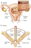
FIGURE 2. Comparative anatomy of the uterine circulation in humans, rodents, and ungulates
In most mammals, including humans (A), blood is delivered to the uterus bidirectionally via a dual arterial anastomotic loop in which one end (the two ovarian arteries) originates from the aorta and the other (uterine arteries) from the internal iliacs. Unlike the linear, branching pattern of many vascular networks in which blockage leads to downstream ischemia, this bilateral anatomical arrangement provides the uterus with a dual source of blood and considerable redundancy in case of occlusion. Perpendicular vessels arise from the main utero-ovarian arteries and pass into the body of the uterus to form the arcuate arteries, which encircle the organ by coursing within the myometrium just beneath its serosal (outer) surface; vessels from each side anastomose along the uterine midline. Smaller radial arteries (see inset) emanate from the arcuates and penetrate the myometrium centripetally before ramifying into either straight (basal) or coiled (spiral) arteries at the myoendometrial border. The basal vessels spread to form a network along the myoendometrial border, while the spiral arteries penetrate further into the endometrium and terminate close to the uterine lumen in capillaries that are, in turn, drained by venules that coalesce into larger veins that eventually enter the inferior vena cava (not shown). Rodents (B) have a duplex uterus, with the main utero-ovarian (or parametrial) arteries and veins running parallel to, but well outside of, the uterine wall within a planar sheet of connective tissue called the mesometrium (shown by tan shading). The vessels of the mesometrium are perfused by arterial blood coming from either the uterine or the ovarian end, i.e., with bidirectional flow. Secondary vessels analogous to the arcuate arteries in humans may form redundant loops with the main artery, and tertiary radial arteries connect the arcuate loops with the uterine wall. These radial (also called mesometrial or segmental) arteries can be further categorized based on their destination as being either premyometrial or preplacental. The premyometrial radial arteries enter the uterine wall between placentation sites and ramify into an intrauterine arterial plexus that supplies the myometrium, whereas preplacental radial vessels widen before entering the placenta through a process of endovascular trophoblast invasion. In addition to displaying distinctive patterns of remodeling during gestation, premyometrial vs. preplacental arteries have also been shown to have different patterns of reactivity (see text). In ungulates such as the sheep or pig, the main (middle) uterine artery originates from the umbilical branch of the internal iliac artery and divides into four primary branches that anastomose with contralateral vessels along the lesser curvature of the uterine horn. These vessels give rise to coiled, branching vessels that run along the ventral and dorsal surface of the uterus to form the arcuate arteries, with smaller branches (radial arteries) that penetrate the myometrium and terminate in arterioles within the endometrium. Most notably, whereas humans and rodents exhibit a hemochorial type of placentation (having low resistance), the placenta of sheep and pigs is epitheliochorial and therefore more analogous to a true microcirculatory bed. In humans, as well as other species (e.g., sheep, pig, rat, guinea pig), the uterus is drained by a venous system that parallels the arterial tree, with closely apposed arteries and veins.
FIGURE 3
FIGURE 3. Preplacental vs. premyometrial vessels in the rat
Photograph of a segment of the uterine mesometrium from a 20-day pregnant rat. A proximal radial artery (RA) divides into two branches, both of which approach the uterine wall (not visible). The lower vessel perfuses the placenta [preplacental artery (PPL)] while the upper courses to and penetrates the myometrium between implantation sites [premyometrial (PMM)]. A segment of a vein draining the placenta is visible at top right. Note consistent shape of the PMM vs. significant (2–3×) widening of the PPL. The area of widening likely denotes the extent of endovascular trophoblast invasion. Once the vessel has widened, it loses the ability to contract or dilate due to ablation and/or de-differentiation of vascular smooth muscle in the arterial media.
FIGURE 4
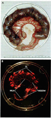
FIGURE 4. Local mechanisms predominate in uterine vascular remodeling in the rodent
A: photograph of a 20-day pregnant rat uterus in which one uterine horn was ligated at the ovarian end, preventing oocyte descent and fertilization. Thirteen fetoplacental units are present in the pregnant uterine horn (PH); the nonpregnant horn is at the bottom of the picture (NPH). CERV, cervix. B: the arterial vasculature of the specimen in A was infused with a latex casting compound and allowed to harden. Uterine and fetoplacental tissues were digested in KOH, leaving a cast of the arterial circulation of each horn. The main uterine arteries (MUA) and the mesometrial arteries (MESOM; arcuate, radial) are labeled, as are two small tree-like structures that represent the maternal intraplacental compartment typical of rodent hemochorial placentation (PL).
FIGURE 5
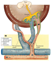
FIGURE 5. Venoarterial exchange
Signals secreted from the placenta, decidua, or myometrium (due to stretch) pass into the venous effluent, where their concentrations would be highest before dilution into the systemic circulation. Signals may be molecules that are vasoactive, mitogenic, or hypertrophic. These, in turn, pass across the venous wall to the adjacent artery to alter arterial tone and/or structure. This pathway could provide a mechanism for fetoplacental regulation of maternal blood flow. Although venoarterial exchange has been documented in the uterine circulation of a number of species as a mechanism for luteolysis (see text), its role in maternal uterine vascular remodeling during pregnancy is still hypothetical.
FIGURE 6
FIGURE 6. Four physiological mechanisms that may play a role in uterine arterial and venous widening and elongation during pregnancy
Note: This generalized drawing of the uteroplacental circulation with hemochorial placentation is not meant to be anatomically accurate; rather, it summarizes the four principal mechanisms likely to be involved in gestational uterine vascular remodeling, as discussed in the text. 1) Placentation/endovascular trophoblast invasion. Hemochorial placentation provides a low-resistance pathway for maternal blood flow by eliminating the intramyometrial microcirculation and creating an intervillous space (IVS). Flow resistance is further decreased by arterial widening secondary to endovascular trophoblast invasion (green cells) of the preplacental (in humans, spiral) arteries. Invasion of the veins has also been documented, at least in rodents, although to a lesser depth. The depth of invasion is exaggerated for purposes of illustration. 2) Increased shear stress. Decreased resistance due to placentation and endovascular remodeling of distal (preplacental) arteries results in an acceleration of blood flow (Q) in proximal vessels, elevating shear stress at the endothelial surface (2, black arrows). Shear stimulates the release of endothelial NO and initiates outward circumferential (expansive) arterial remodeling. As vessels enlarge, higher flow can be maintained at a slower velocity, thereby normalizing shear stress at the endothelial surface. Venous shear stress must increase as well (since inflow must equal outflow), and venous circumferential enlargement and elongation clearly occur, although a role for shear stress in venous expansive remodeling has not yet been demonstrated experimentally. 3) Venoarterial exchange. 3a: placentally derived signals. The fetoplacental unit secretes a variety of signals (blue circles) into the IVS. These pass into the venous outflow, where they are most concentrated before systemic dilution and may 1) induce changes in venous structure and 2) enhance venous permeability through molecular mechanisms (e.g., VEGF, PIGF) and altered physical forces secondary to growth (e.g., increased wall tension). Both mechanisms have been described in the uterine circulation of rodents (see text), where arteries and veins are often in close apposition and would affect vessels within the area shown by the tan shaded box and indicated by the placement of symbols between the arteries and veins. Placental signals may include molecules that reduce arterial tone and stimulate cell division and/or hypertrophy, leading to vessel widening. 3b: myometrial signals. Mechanisms involved in the axial remodeling (elongation) of arteries and veins have not been identified to date. Here, we postulate that increasing volume of the conceptus (white arrows) leads to myometrial stretch. This, in turn, induces the release of mitogenic signals (black triangles) into the venous outflow, stimulating hyperplasia within the venous wall (and in the adjacent arterial wall via venoarterial exchange) and leading to structural elongation. Human arteries must also elongate during pregnancy, since the volume of the uterus increases approximately 1,000-fold, although it is not clear whether lengthening is due to arterial growth, elastic stretch, straightening of tortuous segments, or any combination thereof. In rodents, much of the vasculature is external to the myometrium and located in a planar mesometrium; in humans, arcuate and radial arteries are contained within the myometrium and may therefore be subject to direct stretch. 4) Humoral factors. The endocrine milieu of pregnancy results in altered systemic concentrations of hormones and growth factors (red circles). The source of hormones may be placental, although this is species dependent (e.g., in rodents, the ovary, not the placenta, produces estrogen and progesterone). Increased hormone concentrations may induce vasodilation, alter permeability, and stimulate both cellular and matrix remodeling.

Similar articles

Cited by

References

    1. Adamson SL, Lu Y, Whiteley KJ, Holmyard D, Hemberger M, Pfarrer C, Cross JC. Interactions between trophoblast cells and the maternal and fetal circulation in the mouse placenta. Dev Biol. 2002;250:358–373. - PubMed
    1. Annibale DJ, Rosenfeld CR, Stull JT, Kamm KE. Protein content and myosin light chain phosphorylation in uterine arteries during pregnancy. Am J Physiol Cell Physiol. 1990;259:C484–C489. - PubMed
    1. Assali NS, Douglass RA, Jr, Baird WW, Nicholson DB, Suyemoto R. Measurement of uterine blood flow and uterine metabolism. IV. Results in normal pregnancy. Am J Obstet Gynecol. 1953;66:248–253. - PubMed
    1. Assali NS, Rauramo L, Peltonen T. Measurement of uterine blood flow and uterine metabolism VIII. Uterine and fetal blood flow and oxygen consumption in early human pregnancy. Am J Obstet Gynecol. 1960;79:86–98. - PubMed
    1. Ben Driss A, Benessiano J, Poitevin P, Levy BI, Michel JB. Arterial expansive remodeling induced by high flow rates. Am J Physiol Heart Circ Physiol. 1997;272:H851–H858. - PubMed

Publication types

LinkOut - more resources