. 2009 Feb 15;23(4):452-65.
doi: 10.1101/gad.1749309. Epub 2009 Feb 4.

Linker histone H1 is essential for Drosophila development, the establishment of pericentric heterochromatin, and a normal polytene chromosome structure

Affiliations

Linker histone H1 is essential for Drosophila development, the establishment of pericentric heterochromatin, and a normal polytene chromosome structure

Xingwu Lu et al. Genes Dev. .

Abstract

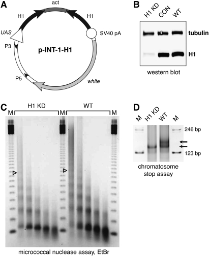
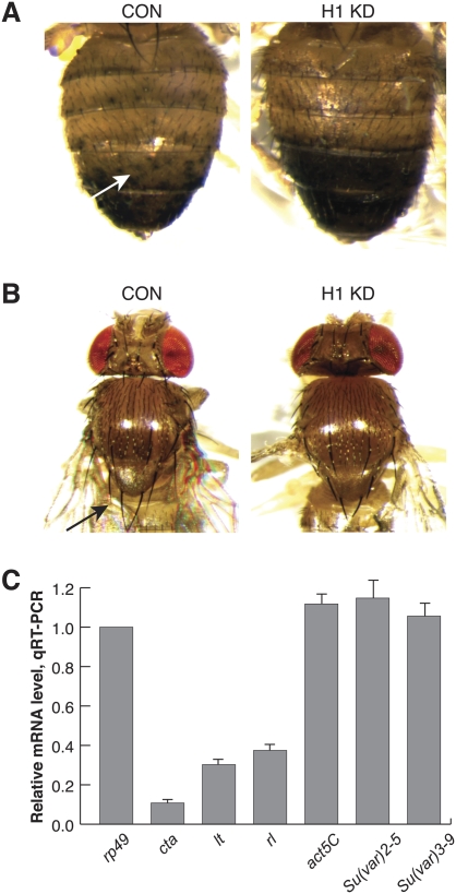
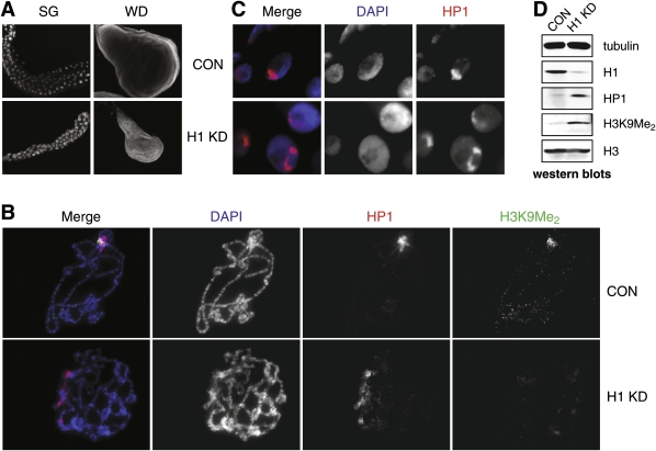
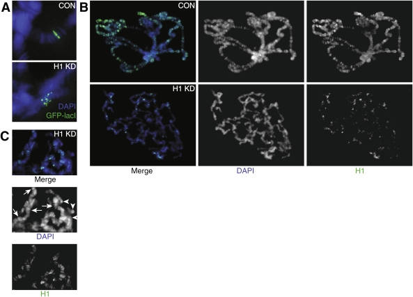
We generated mutant alleles of Drosophila melanogaster in which expression of the linker histone H1 can be down-regulated over a wide range by RNAi. When the H1 protein level is reduced to approximately 20% of the level in wild-type larvae, lethality occurs in the late larval - pupal stages of development. Here we show that H1 has an important function in gene regulation within or near heterochromatin. It is a strong dominant suppressor of position effect variegation (PEV). Similar to other suppressors of PEV, H1 is simultaneously involved in both the repression of euchromatic genes brought to the vicinity of pericentric heterochromatin and the activation of heterochromatic genes that depend on their pericentric localization for maximal transcriptional activity. Studies of H1-depleted salivary gland polytene chromosomes show that H1 participates in several fundamental aspects of chromosome structure and function. First, H1 is required for heterochromatin structural integrity and the deposition or maintenance of major pericentric heterochromatin-associated histone marks, including H3K9Me(2) and H4K20Me(2). Second, H1 also plays an unexpected role in the alignment of endoreplicated sister chromatids. Finally, H1 is essential for organization of pericentric regions of all polytene chromosomes into a single chromocenter. Thus, linker histone H1 is essential in Drosophila and plays a fundamental role in the architecture and activity of chromosomes in vivo.


Figures

Figure 1.
Figure 1.
Depletion of histone H1 in Drosophila. (A) Transgenic RNAi construct for sequence-specific post-transcriptional silencing of the Drosophila linker histone H1 gene. Two identical fragments of the H1 coding sequence (the first 600 bp) were inserted in opposite orientations on both sides of the first intron of the actin 5C gene in the pINT-1 vector. The expression of H1-specific dsRNA is driven by the GAL4-responsive UAS promoter. (Open triangle) UAS promoter; (black arrows) H1 cDNA fragments; (dark-gray box) act5C intron; (open circle) SV40 polyadenylation site; (light arrow) white gene; (small black arrowheads) P-element sequences. (B) The expression of H1 protein in the third instar (L3) larval salivary glands is abrogated by RNAi. H1 synthesis was inhibited by H1-specific dsRNA transcribed from p-INT-1-H15M transgene driven by the GAL4 transactivator encoded by the Tubulin-GAL4 transgene at 29°C (from the beginning of embryonic development to L3). H1 protein levels in salivary gland lysates of H1-depleted animals (H1 KD) were compared with those in salivary glands of the wild-type (WT) and pINT-1-Nautilus (CON) animals by immunoblotting using an antiserum against Drosophila histone H1. Protein loading was controlled by immunoblotting for tubulin. (C) Nucleosome repeat length (NRL) is reduced in L3 larval chromatin upon depletion of H1 by RNAi. H1 synthesis was inhibited by H1-specific dsRNA as described in B. Nuclei from L3 larvae were subjected to partial micrococcal nuclease digestion, and the DNA was analyzed by agarose gel electrophoresis and EtBr staining. The NRL in knockdown animals (H1 KD) was calculated to be ∼176 bp compared with ∼188 bp in the wild-type control (WT). Open triangles indicate the positions of the hexanucleosome bands in each sample. (M) 123-bp DNA ladder. (D) Chromatosome particles are depleted in the chromatin of H1 knockdown larvae. Chromatin from H1 knockdown (H1 KD) and wild-type (WT) larvae was prepared and digested extensively with MNase as in C. The DNA was analyzed by native PAGE and EtBr staining. In H1 knockdown larvae, the chromatosome band (top arrow) is substantially depleted in comparison with the wild-type control larvae. (Bottom arrow) Core particle; (M) 123-bp DNA ladder.
Figure 2.
Figure 2.
H1 is a suppressor of PEV and an activator of heterochromatic gene expression. (A) Variegation of a yellow transgene transposed into pericentric heterochromatin in 2R is suppressed by partial depletion of H1. The recessive yellow gene within the SUPorP P-element is inserted into pericentric heterochromatin (KV111, 2h46) (Konev et al. 2003). (Left panel) Variegated expression of yellow manifests in the dorsal abdominal cuticle as dispersed patches of cells with or without the dark pigment (arrow). (Right panel) In H1-depleted adult flies (pINT-1-H12M + Tubulin-GAL4), variegation of yellow is suppressed, which is indicated by intensification of the dark pigment in the dorsal abdomen. The animals were incubated at 22°C throughout their life cycle. (H1 KD) H1-depleted animals; (CON) Nautilus RNAi control. (B) Variegation of Sb[V] allele in a rearranged Chromosome 3, T(2;3)Sb[V], is suppressed by partial depletion of H1. (Left panel) Silencing of heterochromatin-proximal Sb[V] causes a variegated increase of bristle length toward the wild-type phenotype (arrow). (Right panel) H1 depletion (pINT-1-H12M + Tubulin-GAL4) causes a suppression of PEV, which is indicated by an increased number of short bristles. The animals were incubated at 22°C throughout their life cycle. (H1 KD) H1-depleted animals; (CON) Nautilus RNAi control. (C) H1 depletion causes repression of pericentric genes concertina, light, and rolled. Total RNA was prepared from dissected salivary glands of H1-depleted (pINT-1-H15M + Tubulin-GAL4) and control (pINT-1-Nau + Tubulin-GAL4) L3 larvae, and mRNA levels were analyzed in triplicate by real-time qRT–PCR. The measurements were normalized to rp49. Gene expression levels in H1-depleted larvae are expressed relative to the Nautilus RNAi control. The animals were incubated at 29°C throughout their life cycle. act5C, Su(var)2-5, and Su(var)3-9 gene expression levels were measured as controls.
Figure 3.
Figure 3.
H1 depletion affects HP1 localization and the status of H3K9 dimethylation in Drosophila polytene chromosomes. (A) H1 depletion by RNAi leads to a reduction in the number of cells and the size of salivary glands and imaginal discs in L3 larvae. Whole salivary glands and wing imaginal discs were dissected from H1-depleted (pINT-1-H15M + Tubulin-GAL4) and control (pINT-1-Nau + Tubulin-GAL4) L3 larvae. The animals were incubated at 29°C throughout their life cycle. Tissues were fixed and stained with DAPI to visualize the nuclei. (SG) Salivary gland; (WD) wing disc; (CON) pINT-1-Nau + Tubulin-GAL4 control larvae; (H1 KD) pINT-1-H15M + Tubulin-GAL4 H1 RNAi larvae. (B) H1 depletion causes abnormal polytene chromosome morphology and the loss of the H3K9Me2 marker in pericentric heterochromatin. Salivary glands from control (pINT-1-Nau + Tubulin-GAL4) and H1-depleted (pINT-1-H15M + Tubulin-GAL4) L3 larvae were squashed, and polytene spreads were stained with DAPI and antibodies against HP1 and H3K9Me2. Control chromosomes (CON) have a uniform regular structure of bands and interbands as well as a prominent chromocenter brightly stained with DAPI and both antibodies, whereas no such uniform banding pattern or the chromocenter are apparent in the H1-depleted polytene chromosomes (H1 KD). In contrast to the control chromosomes, the H1-depleted polytene chromosome structure is severely disturbed, HP1 is dispersed broadly, and the H3K9Me2 cannot be readily detected, including at the loci where HP1 is present. The animals were incubated at 29°C throughout their life cycle. (Blue) DAPI; (red) HP1; (green) H3K9Me2. (C) HP1 is distributed in more than one chromosomal locus in salivary gland cells with depleted H1. Whole-mount salivary glands from control (pINT-1-Nau + Tubulin-GAL4, CON) and H1-depleted (pINT-1-H15M + Tubulin-GAL4, H1 KD) larvae were fixed and stained with anti-HP1 antibody. Whereas in control cells HP1 is mostly concentrated in a single region (chromocenter), in H1-depleted cells HP1 is dispersed broadly. The animals were incubated at 29°C throughout their life cycle. (Blue) DAPI; (red) HP1. (D) Total cellular HP1 and H3K9Me2 are increased upon abrogation of H1 expression in larvae. HP1 and H3K9Me2 were detected by Western blotting of crude lysates of salivary glands from control (pINT-1-Nau + Tubulin-GAL4, CON) and H1-depleted (pINT-1-H15M + Tubulin-GAL4, H1 KD) L3 larvae. Western blotting for tubulin and total histone H3 were used as loading controls. The animals were incubated at 29°C throughout their life cycle.
Figure 4.
Figure 4.
Effects of H1 depletion on alignment of sister chromatids in polytene chromosomes. (A) H1 depletion results in misalignment of chromatin fibrils in an interband region in 61F. The genomic position of a P-element insertion containing 256 lacO sites was visualized by indirect immunofluorescence (IF) through binding of an ectopically expressed GFP-lacI protein to the locus in polytene chromosomes. In control squashes (pINT-1-Nau + Tubulin-GAL4, CON), the GFP signal was observed as a single straight band, indicative of an alignment of endoreplicated chromatin fibrils. In H1-depleted polytene chromosomes (pINT-1-H15M + Tubulin-GAL4, H1 KD), the GFP signal was dispersed into multiple isolated spots, indicating the loss of perfect alignment. The animals were incubated at 29°C throughout their life cycle. (Blue) DAPI; (green) GFP-lacI. (B) Residual H1 protein in H1-depleted polytene chromosomes is unevenly distributed and correlates with regions of persistent polytene band–interband structure. Polytene chromosome squashes from control (pINT-1-Nau + Tubulin-GAL4, CON) and H1-depleted (pINT-1-H15M + Tubulin-GAL4, H1 KD) salivary glands were stained with the H1 antibody. In control chromosomes, H1 localizes primarily to bands in euchromatic arms and to the pericentric region. In H1-depleted larvae, the H1 signal is dramatically reduced, and the residual protein is distributed over a limited number of loci. The majority of these loci also have a partially conserved polytene band–interband structure, as evidenced by DAPI staining. In contrast, chromosome regions that do not contain detectable H1 typically exhibit amorphous, clumped morphology. The animals were incubated at 29°C throughout their life cycle. (Blue) DAPI; (green) H1. (C) In H1 knockdown larvae, polytene band–interband structure is partially preserved in loci with elevated residual H1. Whereas DAPI stains equally brightly the residual structured bands (arrows) and unstructured chromatin clumps (arrowheads), H1 staining predominantly has an appearance of discrete bands. No diffuse signal can be observed for the H1 staining. Furthermore, there is a substantial overlap between the residual DAPI and H1 bands.

References

    1. Ananiev E.V., Barsky V.E. Elementary structures in polytene chromosomes of Drosophila melanogaster. Chromosoma. 1985;93:104–112. - PubMed
    1. Bao X., Deng H., Johansen J., Girton J., Johansen K.M. Loss-of-function alleles of the JIL-1 histone H3S10 kinase enhance position-effect variegation at pericentric sites in Drosophila heterochromatin. Genetics. 2007;176:1355–1358. - PMC - PubMed
    1. Brand A.H., Manoukian A.S., Perrimon N. Ectopic expression in Drosophila. Methods Cell Biol. 1994;44:635–654. - PubMed
    1. Brown D.T., Alexander B.T., Sittman D.B. Differential effect of H1 variant overexpression on cell cycle progression and gene expression. Nucleic Acids Res. 1996;24:486–493. - PMC - PubMed
    1. Cartwright I.L., Cryderman D.E., Gilmour D.S., Pile L.A., Wallrath L.L., Weber J.A., Elgin S.C. Analysis of Drosophila chromatin structure in vivo. Methods Enzymol. 1999;304:462–496. - PubMed

Publication types

MeSH terms

LinkOut - more resources