

Proliferation of uterine natural killer cells is induced by human chorionic gonadotropin and mediated via the mannose receptor
- PMID: 19196802
- PMCID: PMC2709965
- DOI: 10.1210/en.2008-1309
Proliferation of uterine natural killer cells is induced by human chorionic gonadotropin and mediated via the mannose receptor
Abstract
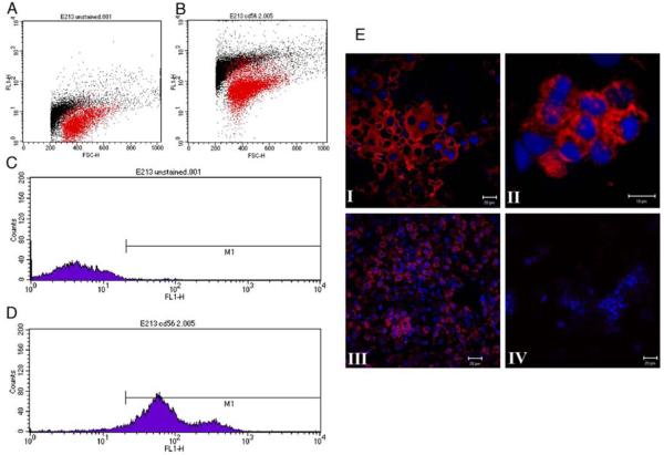
The endometrial lining of the human uterus contains a population of phenotypically distinct (CD56(bright), CD16(dim)), tissue-specific, natural killer [uterine natural killer (uNK)] cells that play a key role in the establishment of a successful pregnancy. An increase in the number of endometrial uNK cells occurs when the conceptus implants, and there is a further increase during the early stages of placentation. Here, we describe studies that have identified human chorionic gonadotrophin (hCG), a glycoprotein synthesized by the preimplantation conceptus, as a novel regulator of uNK cell proliferation. The impact of hCG on uNK cells was mediated via the mannose receptor (CD206) rather than by the classical hCG/LH receptor that was not expressed. The mannose receptor and hCG were colocalized on the surface of uNK cells, and proliferation did not occur if cells were incubated with deglycosylated hCG or intact hCG in the presence of excess d-Mannose. These novel observations provide new insight into the endocrine-immune dialogue that exists between the conceptus and immune cells within the receptive endometrium, and have implications for the role of uNK cell-trophoblast interactions and pregnancy outcome.
Figures


Similar articles
-
Profiling the expression and function of oestrogen receptor isoform ER46 in human endometrial tissues and uterine natural killer cells.Hum Reprod. 2020 Mar 27;35(3):641-651. doi: 10.1093/humrep/dez306. Hum Reprod. 2020. PMID: 32108901 Free PMC article.
-
Receptors for non-MHC ligands contribute to uterine natural killer cell activation during pregnancy in mice.Placenta. 2013 Sep;34(9):757-64. doi: 10.1016/j.placenta.2013.06.004. Epub 2013 Jun 24. Placenta. 2013. PMID: 23806179 Free PMC article.
-
Estrogen-dependent regulation of human uterine natural killer cells promotes vascular remodelling via secretion of CCL2.Hum Reprod. 2015 Jun;30(6):1290-301. doi: 10.1093/humrep/dev067. Epub 2015 Mar 27. Hum Reprod. 2015. PMID: 25820695 Free PMC article.
-
The regulation of ovary and conceptus on the uterine natural killer cells during early pregnancy.Reprod Biol Endocrinol. 2017 Sep 6;15(1):73. doi: 10.1186/s12958-017-0290-1. Reprod Biol Endocrinol. 2017. PMID: 28874155 Free PMC article. Review.
-
Natural killer cell-triggered vascular transformation: maternal care before birth?Cell Mol Immunol. 2011 Jan;8(1):1-11. doi: 10.1038/cmi.2010.38. Epub 2010 Aug 16. Cell Mol Immunol. 2011. PMID: 20711229 Free PMC article. Review.
Cited by
-
Meta-analysis of intrauterine hCG perfusion efficacy in recurrent implantation failure as defined by ESHRE guidelines.BMC Pregnancy Childbirth. 2024 Jul 9;24(1):468. doi: 10.1186/s12884-024-06662-1. BMC Pregnancy Childbirth. 2024. PMID: 38982352 Free PMC article.
-
Promoting Roles of Embryonic Signals in Embryo Implantation and Placentation in Cooperation with Endocrine and Immune Systems.Int J Mol Sci. 2020 Mar 10;21(5):1885. doi: 10.3390/ijms21051885. Int J Mol Sci. 2020. PMID: 32164226 Free PMC article. Review.
-
Transcriptome analysis reveals new insights into the modulation of endometrial stromal cell receptive phenotype by embryo-derived signals interleukin-1 and human chorionic gonadotropin: possible involvement in early embryo implantation.PLoS One. 2013 May 22;8(5):e64829. doi: 10.1371/journal.pone.0064829. Print 2013. PLoS One. 2013. PMID: 23717664 Free PMC article.
-
Endocrine factors modulating immune responses in pregnancy.Front Immunol. 2014 May 8;5:196. doi: 10.3389/fimmu.2014.00196. eCollection 2014. Front Immunol. 2014. PMID: 24847324 Free PMC article. Review.
-
Granulocyte colony stimulating factor versus human chorionic gonadotropin for recurrent implantation failure in intra cytoplasmic sperm injection: a randomized clinical trial.BMC Pregnancy Childbirth. 2022 Nov 29;22(1):881. doi: 10.1186/s12884-022-05098-9. BMC Pregnancy Childbirth. 2022. PMID: 36447142 Free PMC article. Clinical Trial.
References
-
- King A. Uterine leukocytes and decidualization. Human Reproduction Update. 2000;6:28–36. - PubMed
-
- Moffett A, Loke C. Immunology of placentation in eutherian mammals. Nat Rev Immunol. 2006;6:584–594. - PubMed
-
- Bulmer JN, Morrison L, Longfellow M, Ritson A, Pace D. Granulated lymphocytes in human endometrium: histochemical and immunohistochemical studies. Hum Reprod. 1991;6:791–798. - PubMed
-
- Dosiou C, Giudice LC. Natural killer cells in pregnancy and recurrent pregnancy loss: endocrine and immunologic perspectives. Endocr Rev. 2005;26:44–62. - PubMed
Publication types
MeSH terms
Substances
Grants and funding
LinkOut - more resources
Full Text Sources
Other Literature Sources
Research Materials
