. 2009 May;37(5):1035-45.
doi: 10.1124/dmd.108.023317. Epub 2009 Feb 5.

Constitutive androstane receptor-mediated changes in bile acid composition contributes to hepatoprotection from lithocholic acid-induced liver injury in mice

Affiliations

Constitutive androstane receptor-mediated changes in bile acid composition contributes to hepatoprotection from lithocholic acid-induced liver injury in mice

Lisa D Beilke et al. Drug Metab Dispos. 2009 May.

Abstract

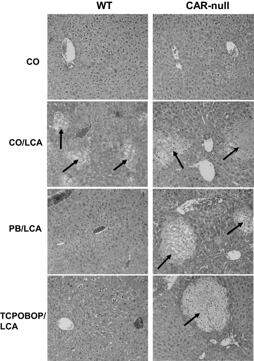
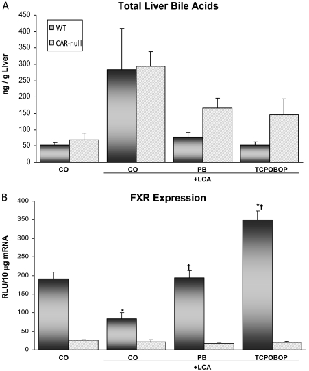
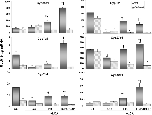
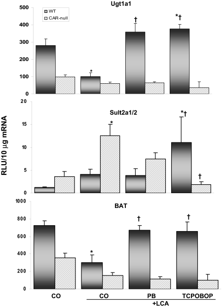
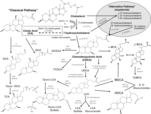
Pharmacological activation of the constitutive androstane receptor (CAR) protects the liver during cholestasis. The current study evaluates how activation of CAR influences genes involved in bile acid biosynthesis as a mechanism of hepatoprotection during bile acid-induced liver injury. CAR activators phenobarbital (PB) and 1,4-bis[2-(3,5-dichloropyridyloxy)]benzene (TCPOBOP) or corn oil (CO) were administered to C57BL/6 wild-type (WT) and CAR knockout (CAR-null) mice before and during induction of intrahepatic cholestasis using the secondary bile acid, lithocholic acid (LCA). In LCA-treated WT and all the CAR-null groups (excluding controls), histology revealed severe multifocal necrosis. This pathology was absent in WT mice pretreated with PB and TCPOBOP, indicating CAR-dependent hepatoprotection. Decreases in total hepatic bile acids and hepatic monohydroxy, dihydroxy, and trihydroxy bile acids in PB- and TCPOBOP-pretreated WT mice correlated with hepatoprotection. In comparison, concentrations of monohydroxylated and dihydroxylated bile acids were increased in all the treated CAR-null mice compared with CO controls. Along with several other enzymes (Cyp7b1, Cyp27a1, Cyp39a1), Cyp8b1 expression was increased in hepatoprotected mice, which could be suggestive of a shift in the bile acid biosynthesis pathway toward the formation of less toxic bile acids. In CAR-null mice, these changes in gene expression were not different among treatment groups. These results suggest CAR mediates a shift in bile acid biosynthesis toward the formation of less toxic bile acids, as well as a decrease in hepatic bile acid concentrations. We propose that these combined CAR-mediated effects may contribute to the hepatoprotection observed during LCA-induced liver injury.


Figures

F<sc>ig</sc>. 1.
Fig. 1.
CAR activators protect the liver against bile acid-induced toxicity. A midsection of the left liver lobe was removed and fixed from each animal. Tissues were stained with hematoxylin and eosin, and histopathology was determined under treatment-blinded conditions by a board-certified veterinary pathologist. Two animals per treatment group were evaluated, and pictures are representative of treatment group pathology. Multifocal hepatic necrosis (arrows) is easily distinguished from surrounding parenchyma and is more extensive in mice treated with LCA; 50× magnification.
F<sc>ig</sc>. 2.
Fig. 2.
Total hepatic bile acid concentrations and FXR expression. Animals (n = 4–6/group) were dosed with activators and LCA (125 mg/kg twice daily) as described under Materials and Methods. A, bile acids were extracted from the liver, and the concentration was determined using 3α-hydroxysteroid dehydrogenase. Liver bile acid concentrations are presented as nanograms per gram of liver tissue. B, total RNA was isolated from livers of WT and CAR-null mice. The data are presented as mean relative light units ± S.E.M. *, indicates p ≤ 0.05 compared with respective CO; †, indicates p ≤ 0.05 compared with respective LCA only.
F<sc>ig</sc>. 3.
Fig. 3.
Hydroxylated bile acid concentrations in the liver. Animals (n = 4–6/group) were pretreated with activators and LCA (125 mg/kg twice daily) as described under Materials and Methods. Results are presented as mean concentration ± S.E.M. *, indicates p ≤ 0.05 compared with respective CO; †, indicates p ≤ 0.05 compared with respective LCA only.
F<sc>ig</sc>. 4.
Fig. 4.
Expression of phase I bile acid biosynthesis and metabolizing genes. Hepatic Cyp3a11, Cyp7a1, Cyp7a1, Cyp8a1, Cyp27a1, and Cyp39a1 mRNA levels in each treatment group were quantified by the bDNA signal amplification assay, as described under Materials and Methods. Data are expressed as relative light units (RLU) ± S.E.M. *, indicates p ≤ 0.05 compared with respective CO; †, indicates p ≤ 0.05 compared with respective LCA only.
F<sc>ig</sc>. 5.
Fig. 5.
Expression of phase II bile acid-conjugating genes. Hepatic Sult2A1/2, Ugt1A1, and BAT mRNA levels in each treatment group were quantified by the bDNA signal amplification assay, as described under Materials and Methods. Data are expressed as relative light units (RLU) ± S.E.M. *, indicates p ≤ 0.05 compared with respective CO; †, indicates p ≤ 0.05 compared with respective LCA-only.
F<sc>ig</sc>. 6.
Fig. 6.
Simplified overview of the bile acid biosynthesis pathway derived from cholesterol. The neutral (classic) and acidic (alternative) routes are shown with the main enzymes involved. The 7α-hydroxylation of cholesterol by CYP7A1 is the rate-limiting enzyme in the neutral pathway. The neutral pathway is considered the most important pathway for bile acid formation in humans, whereas the acidic pathway is important for the removal of cholesterol from extrahepatic tissues, and it seems to be able to compensate for the neutral pathway when it is repressed to maintain bile acid formation. Boxed area highlights the shift in biosynthesis via up-regulation of Cyp8b1 in hepatoprotected mice. (Note: not all the bile acids, enzymes, or intermediate steps shown.) Chemical structure sources: http://sigmaaldrich.com, http://steraloids.com.

References

    1. Alnouti Y and Klaassen CD (2006) Tissue distribution and ontogeny of sulfotransferase enzymes in mice. Toxicol Sci 93 242–255. - PubMed
    1. Annapurna V, Mukundan M, Sesikeran B, and Bamji M (1989) Effects of ovulen-50, diethylnitrosamine and phenobarbital on liver regeneration in female rats. J Biosci 14 1–7.
    1. Araya Z and Wikvall K (1999) 6alpha-Hydroxylation of taurochenodeoxycholic acid and lithocholic acid by CYP3A4 in human liver microsomes. Biochim Biophys Acta 1438 47–54. - PubMed
    1. Assem M, Schuetz EG, Leggas M, Sun D, Yasuda K, Reid G, Zelcer N, Adachi M, Strom S, Evans RM, et al. (2004) Interactions between hepatic Mrp4 and SULT2a1/2 as revealed by the constitutive androstane receptor and Mrp4 knockout mice. J Biol Chem 279 22250–22257. - PubMed
    1. Baskin-Bey E, Anan A, Isomoto H, Bronk S, and Gores G (2007) Constitutive androstane receptor agonist, TCPOBOP, attenuates steatohepatitis in the methionine choline-deficient diet-fed mouse. World J Gastroenterol 13 5635–5641. - PMC - PubMed

Publication types

MeSH terms