. 2009 Feb 24;106(8):2770-5.
doi: 10.1073/pnas.0807694106. Epub 2009 Feb 5.

Absence of autophagy results in reactive oxygen species-dependent amplification of RLR signaling

Affiliations

Absence of autophagy results in reactive oxygen species-dependent amplification of RLR signaling

Michal Caspi Tal et al. Proc Natl Acad Sci U S A. .

Abstract

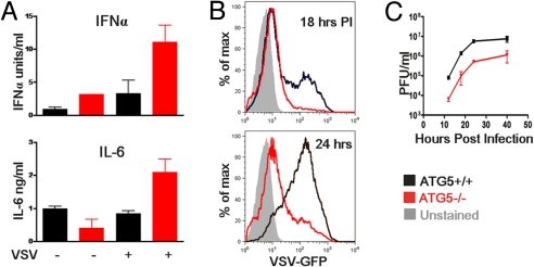
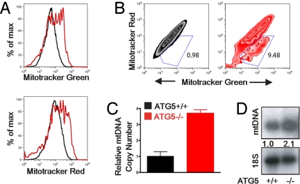
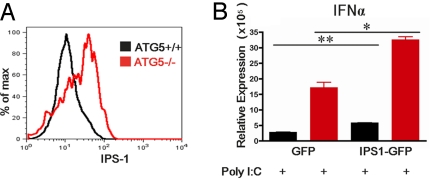
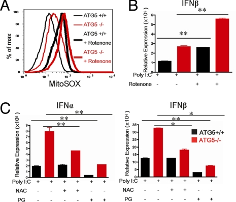
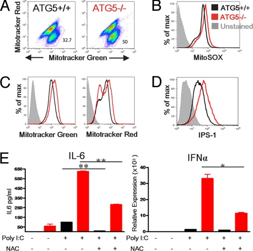
Autophagy is a highly conserved process that maintains homeostasis by clearing damaged organelles and long-lived proteins. The consequences of deficiency in autophagy manifest in a variety of pathological states including neurodegenerative diseases, inflammatory disorders, and cancer. Here, we studied the role of autophagy in the homeostatic regulation of innate antiviral defense. Single-stranded RNA viruses are recognized by the members of the RIG-I-like receptors (RLRs) in the cytosol. RLRs signal through IPS-1, resulting in the production of the key antiviral cytokines, type I IFNs. Autophagy-defective Atg5(-/-) cells exhibited enhanced RLR signaling, increased IFN secretion, and resistance to infection by vesicular stomatitis virus. In the absence of autophagy, cells accumulated dysfunctional mitochondria, as well as mitochondria-associated IPS-1. Reactive oxygen species (ROS) associated with the dysfunctional mitochondria were largely responsible for the enhanced RLR signaling in Atg5(-/-) cells, as antioxidant treatment blocked the excess RLR signaling. In addition, autophagy-independent increase in mitochondrial ROS by treatment of cells with rotenone was sufficient to amplify RLR signaling in WT cells. These data indicate that autophagy contributes to homeostatic regulation of innate antiviral defense through the clearance of dysfunctional mitochondria, and revealed that ROS associated with mitochondria play a key role in potentiating RLR signaling.


Conflict of interest statement

The authors declare no conflict of interest.

Figures

Fig. 1.
Fig. 1.
Atg5-deficient MEFs show increased cytokine production to Poly I:C and VSV stimulation and are resistant to infection. WT, and Atg5−/− MEFs were incubated with VSV-GFP (at a multiplicity of infection of 4) or transfected with 1 μg/mL Poly I:C. Twelve hours later, IFNα and IL-6 production was assessed by ELISA (A). WT and Atg5−/− MEFs were infected with VSV-GFP (at a multiplicity of infection of 1) and the levels of infection were determined by measuring GFP expression by FACS at 18 and 24 h after infection (B) and by measuring viral titers in the supernatants by plaque assay at the indicated time points (C). Results are representative of 6 separate experiments.
Fig. 2.
Fig. 2.
Dysfunctional mitochondria accumulate in Atg5-deficient MEFs. (A and B) Atg5+/+ and Atg5−/− MEFs were stained with 100 nM MitoTracker Green (which stains the lipid membrane of the mitochondria) and 100 nM MitoTracker Red (which fluoresces upon oxidation in respiring mitochondria). Histograms (A) and contour plots (B) of FACS analysis are depicted. (C) Mitochondrial DNA copy number was measured by quantitative PCR and normalized to nuclear DNA levels in a ratio of mtDNA COI over 18S rDNA. Relative mitochondrial DNA copy numbers are depicted. (D) Mitochondrial DNA levels were assessed by Southern blotting using a mitochondrial DNA-specific probe. Numbers indicate relative intensity of mtDNA normalized to cellular DNA. Data are representative of 3 similar experiments.
Fig. 3.
Fig. 3.
IPS-1 overexpression in Atg5−/− MEFs. (A) Intracellular staining of IPS-1 protein analyzed by FACS in Atg5+/+ and Atg5−/− MEFs. (B) Atg5+/+ and Atg5−/− MEFs were transduced with lentivirus expressing IPS-1-GFP fusion protein or GFP (control). Cells expressing only high levels of GFP were sorted by FACS and stimulated with 1 μg/mL Poly I:C complexed to Lipofectamine. Twelve hours later, IFN-α and IFN-β mRNA levels were assessed by RT quantitative PCR. *P < 0.05, **P < 0.005. Similar results were obtained from 2 separate experiments.
Fig. 4.
Fig. 4.
Autophagy-deficient MEFs have increased levels of mitochondrial ROS. Atg5+/+ and Atg5−/− MEFs treated with 1 μM of rotenone for 12 h. (A) Levels of mitochondria associated ROS in Atg5+/+ and Atg5−/− MEFs were analyzed by MitoSOX labeling. (B) After 12 h rotenone pretreatment, cells were transfected with 1 μg/mL Poly I:C. Expression levels of IFN-β were assessed by RT quantitative PCR 12 h after transfection. (C) MEFs were pretreated with 10 mM of the anti-oxidant NAC or 100 μM of PG for 15 min and transfected with Poly I:C (1 μg/mL). IFN-α and IFN-β production was assessed by RT quantitative PCR at 12 h after transfection. *P < 0.05, **P < 0.005. Data are representative of 3 separate experiments.
Fig. 5.
Fig. 5.
Atg5-deficient primary macrophages exhibit amplified RLR signaling. Neonatal liver macrophages were generated from Atg5+/+ or Atg5−/− pups. Cells were stained with 100 nM MitoTracker Green and 100 nM MitoTracker Red as in Fig. 2, and both dot plots (A) and histograms (C) of FACS analysis are depicted. Additionally, cells were labeled with MitoSOX as in Fig. 4 (B) or with antibody to IPS-1 as in Fig. 3. (D) Primary macrophages were transfected with 10 μg/mL Poly I:C in the presence or absence of NAC, and 12 h later, the levels of IL-6 were assessed by ELISA and the levels of IFNα were assessed by RT quantitative PCR (E). *P < 0.05, **P < 0.005. Results are representative of 2 similar experiments.

References

    1. Klionsky DJ, Emr SD. Autophagy as a regulated pathway of cellular degradation. Science. 2000;290:1717–1721. - PMC - PubMed
    1. Mizushima N, Klionsky DJ. Protein turnover via autophagy: implications for metabolism. Annu Rev Nutr. 2007;27:19–40. - PubMed
    1. Levine B, Deretic V. Unveiling the roles of autophagy in innate and adaptive immunity. Nat Rev Immunol. 2007;7:767–777. - PMC - PubMed
    1. Schmid D, Munz C. Innate and adaptive immunity through autophagy. Immunity. 2007;27:11–21. - PMC - PubMed
    1. Pichlmair A, Reis e Sousa C. Innate recognition of viruses. Immunity. 2007;27:370–383. - PubMed

Publication types