. 2009 Feb 24;106(8):2880-5.
doi: 10.1073/pnas.0810530106. Epub 2009 Feb 5.

Structure of a trimeric variant of the Epstein-Barr virus glycoprotein B

Affiliations

Structure of a trimeric variant of the Epstein-Barr virus glycoprotein B

Marija Backovic et al. Proc Natl Acad Sci U S A. .

Abstract

Epstein-Barr virus (EBV) is a herpesvirus that is associated with development of malignancies of lymphoid tissue. EBV infections are life-long and occur in >90% of the population. Herpesviruses enter host cells in a process that involves fusion of viral and cellular membranes. The fusion apparatus is comprised of envelope glycoprotein B (gB) and a heterodimeric complex made of glycoproteins H and L. Glycoprotein B is the most conserved envelope glycoprotein in human herpesviruses, and the structure of gB from Herpes simplex virus 1 (HSV-1) is available. Here, we report the crystal structure of the secreted EBV gB ectodomain, which forms 16-nm long spike-like trimers, structurally homologous to the postfusion trimers of the fusion protein G of vesicular stomatitis virus (VSV). Comparative structural analyses of EBV gB and VSV G, which has been solved in its pre and postfusion states, shed light on gB residues that may be involved in conformational changes and membrane fusion. Also, the EBV gB structure reveals that, despite the high sequence conservation of gB in herpesviruses, the relative orientations of individual domains, the surface charge distributions, and the structural details of EBV gB differ from the HSV-1 protein, indicating regions and residues that may have important roles in virus-specific entry.


Conflict of interest statement

The authors declare no conflict of interest.

Figures

Fig. 1.
Fig. 1.
Structure of the EBV gB Ectodomain. (A) The gB subunit is colored in blue to red, from N and to C terminus, which are labeled N and C, respectively. Numbers indicate N-glycosylated Asn residues, for which a single NAG molecule is modeled. FLs are marked and point in the same direction as the gB C terminus. The ectodomain shown here extends into a fairly hydrophobic, ≈40-residues long, stem region, and a single transmembrane domain, which were removed from the construct used for expression of the recombinant gB due to their high hydrophobicity. The 5 domains of EBV gB are indicated with roman numbers I to V, and are defined and labeled after nomenclature established for the HSV-1 gB ectodomain (Fig. S2 and Table S3). (B) The trimeric gB ectodomain is shown. The subunits are colored as magenta, cyan, and green. The subunit shown on A corresponds to the molecule shown in magenta. The subunits wrap around each other, forming extensive trimerization surfaces.
Fig. 2.
Fig. 2.
Comparison of the EBV and HSV-1 gB Ectodomains. (A) Surface representations of EBV and HSV-1 gB trimers. The most obvious difference in the overall conformation of EBV and HSV-1 gB is the location of domain IV. The domains I and II of the subunit colored in magenta were superimposed and used as a reference point (they occupy the same position in both models). The domains IV assume different locations in EBV and HSV-1 trimers, as emphasized by the arrow. (B) Major structural differences between domains I, II, and III are shown based on their superposition of HSV-1 gB (gray) onto EBV gB (cyan) ectodomain. Domains I, II, and III were superimposed as rigid bodies. The green arrow indicates the rotation of domain IV, also illustrated in A. The yellow arrow points to an additional structural difference in strands β7 and β8 (EBV gB residues 144–157), which are shifted forward and down in EBV gB compared with the HSV-1 molecule. (C) Rotation of the upper part of the central αC (domain III) and domain IV, as seen from a point close to the dashed line drawn in A (coloring is the same as in B). (D) Structural differences in domain I: 2 EBV gB subunits, belonging to the same trimer, are shown in cyan and green, and the superimposed HSV-1 gB is shown in gray. The yellow arrow points to the movement of β7 and β8 of one EBV gB subunit (cyan), which allow the packing of the C-terminal αF of another EBV gB subunit (green). Also indicated is Asn-163 and the attached NAG molecule.
Fig. 3.
Fig. 3.
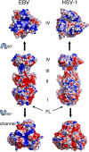
Electrostatic surfaces of EBV and HSV-1 gB ectodomains. (Top) View of domain IV from the top. (Middle) Side view of the trimeric spikes. (Bottom) View of domain I and FLs from below. The views shown at the Top and Bottom are obtained by a 90° rotation from the orientations in the Middle toward and away from the reader, respectively. Despite high conservation of the secondary structure elements and folds of individual domains, electrostatic surfaces of the EBV and HSV-1 gB ectodomains are unique. The electrostatic potential of the EBV trimer tip containing the same 6 FL residues (HR and RVEA) as HSV-1 gB (Bottom) carries no resemblance to the HSV-1 electrostatic landscape, due to contribution of the surrounding EBV residues. The center of the 3 positively charged channels in domain I of EBV gB is indicated by the arrow (Bottom). Electrostatic surfaces were generated by PyMol and are contoured on scale from −2 to +2 kT/e (red corresponds to negative charge, and blue to positive).
Fig. 4.
Fig. 4.
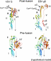
Comparison of VSV G and EBV gB structures. (Upper) Postfusion conformations of VSV G and EBV gB. The structures are colored blue to red from their N to C termini. Domains of gB and G in their postfusion conformations have similar secondary structure topologies, although each of the gB domains is larger, and gB has an additional domain V, which is not present in G. Dashed arrows indicate the missing stem regions, and connect the C termini of the models (marked with letter C) with the expected transmembrane domain position. The prominent αF of VSV G, and the analogous αC of gB, are colored in yellow and indicated with arrows. Disulfide bridges of G, Cys24-Cys284, and gB, Cys68-Cys484, are marked and shown as magenta sticks, connecting the αF and αC, respectively, with the corresponding N-terminal segments (shown in blue). (Lower) The prefusion structure of VSV G and a theoretical model of the prefusion form of EBV gB. In the prefusion conformation of G, only domain III undergoes a significant refolding event (thus, marked as domain III*), which involves breaking of the long αF, whereas the other 3 domains relocate to a different position, but preserve their primary folds. Domain III of gB was modeled by using the domain III* of G as a guide. Dashed arrows represent the direction of stem regions in case of G, and domain V and stem regions for gB.

References

    1. Pellet PE, Roizman B. In: Fields Virology. Knipe DM, Howley PM, editors. New York: Lippincott Williams and Wilkins; 2007. pp. 2479–2499.
    1. Heldwein EE, Krummenacher C. Entry of herpesviruses into mammalian cells. Cell Mol Life Sci. 2008;65:1653–1668. - PMC - PubMed
    1. Spear PG, Longnecker R. Herpesvirus entry: An update. J Virol. 2003;77:10179–10185. - PMC - PubMed
    1. Rey FA. Molecular gymnastics at the herpesvirus surface. EMBO reports. 2006;7:1000–1005. - PMC - PubMed
    1. Haan KM, Kwok WW, Longnecker R, Speck P. Epstein-Barr virus entry utilizing HLA-DP or HLA-DQ as a coreceptor. J Virol. 2000;74:2451–2454. - PMC - PubMed

Publication types

Associated data

LinkOut - more resources