

A role for epithelial-mesenchymal transition in the etiology of benign prostatic hyperplasia
- PMID: 19196965
- PMCID: PMC2650376
- DOI: 10.1073/pnas.0812666106
A role for epithelial-mesenchymal transition in the etiology of benign prostatic hyperplasia
Abstract
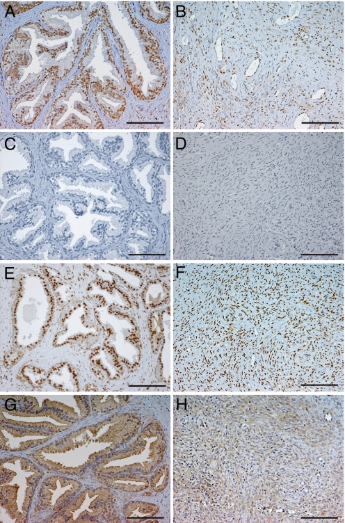
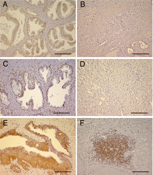
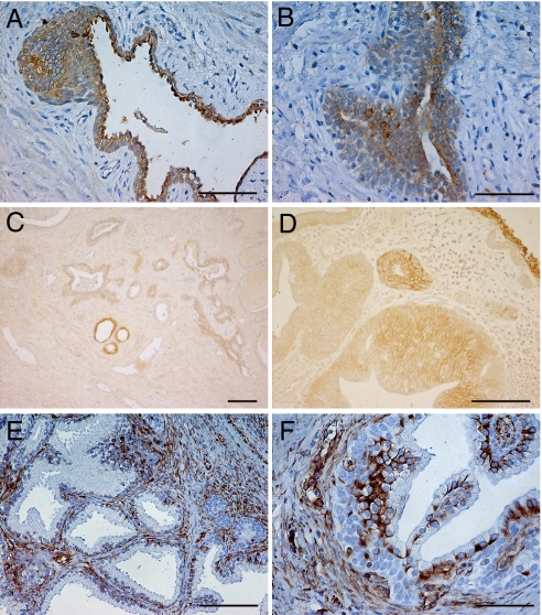
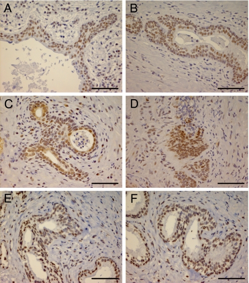
Benign prostatic hyperplasia (BPH) is usually described as a pathological proliferation of prostatic fibroblasts/myofibroblasts and epithelial cells. In the present study of BPH samples, we have made a morphological and immunohistochemical study of BPH prostatic sections using markers of proliferation, apoptosis, hormone receptors, and TGF-beta signaling. We found no evidence of proliferation in the stroma but in the epithelium of some ducts; 0.7% of the basal and 0.4% of luminal cells were positive for Ki67 and PCNA. Androgen receptor and estrogen receptor beta (ERbeta)1 and ERbetacx were abundant in both stromal and epithelial compartments but cells expressing ERalpha were very rare. What was very common in all BPH samples was the following: (i) regions of the ductal epithelium where the epithelial cells did not express E-cadherin, had lost their polarization, and become spindle shaped (the nuclei of these cells were strongly positive for pSmad 3 and Snail); and (ii) regions where the walls of the blood vessels were extremely thick and there was loss of endothelial layer. Loss of E-cadherin, increased pSmad 3, and high expression of Snail are all characteristic of epithelial-mesenchymal transition (EMT). We conclude that BPH is not a disease of prostatic stromal proliferation but rather of accumulation of mesenchymal-like cells derived from the prostatic epithelium and the endothelium. TGF-beta is thought to play a key role in EMT. Our data suggests that TGF-beta/Smad should be considered as targets for treatment of BPH.
Conflict of interest statement
Conflict of interest statement: J.-Å.G. is a shareholder and consultant of KaroBio AB.
Figures
References
-
- Riolan J Lutetiae Parisiorum, editor. Opera Anatomica, Vetera; Recognita and Auctiora. 1649. pp. 874–930.
-
- Rohr HP, Bartsch G. Human benign prostatic hyperplasia: a stromal disease? New perspectives by quantitative morphology. Urology. 1980;16:625–633. - PubMed
-
- Berry SJ, Coffey DS, Walsh PC, Ewing LL. The development of human benign prostatic hyperplasia with age. J Urol. 1984;132:474–479. - PubMed
-
- Tiwari A, Krishna NS, Nanda K, Chugh A. Benign prostatic hyperplasia: an insight into current investigational medical therapies. Expert Opin Investig Drugs. 2005;14:1359–1372. - PubMed
-
- Schroder FH. Medical treatment of benign prostatic hyperplasia: the effect of surgical or medical castration. Prog Clin Biol Res. 1994;386:191–196. - PubMed
Publication types
MeSH terms
Substances
LinkOut - more resources
Full Text Sources
Other Literature Sources
Medical
Research Materials
Miscellaneous
