. 2009 Feb 17;106(7):2097-103.
doi: 10.1073/pnas.0812819106. Epub 2009 Feb 5.

A mouse forward genetics screen identifies LISTERIN as an E3 ubiquitin ligase involved in neurodegeneration

Affiliations

A mouse forward genetics screen identifies LISTERIN as an E3 ubiquitin ligase involved in neurodegeneration

Jessie Chu et al. Proc Natl Acad Sci U S A. .

Abstract

A mouse neurological mutant, lister, was identified through a genome-wide N-ethyl-N-nitrosourea (ENU) mutagenesis screen. Homozygous lister mice exhibit profound early-onset and progressive neurological and motor dysfunction. lister encodes a RING finger protein, LISTERIN, which functions as an E3 ubiquitin ligase in vitro. Although lister is widely expressed in all tissues, motor and sensory neurons and neuronal processes in the brainstem and spinal cord are primarily affected in the mutant. Pathological signs include gliosis, dystrophic neurites, vacuolated mitochondria, and accumulation of soluble hyperphosphorylated tau. Analysis with a different lister allele generated through targeted gene trap insertion reveals LISTERIN is required for embryonic development and confirms that direct perturbation of a LISTERIN-regulated process causes neurodegeneration. The lister mouse uncovers a pathway involved in neurodegeneration and may serves as a model for understanding the molecular mechanisms underlying human neurodegenerative disorders.


Conflict of interest statement

The authors declare no conflict of interest.

Figures

Fig. 1.
Fig. 1.
Progressive impairment of neuronal and motor functions associated with weight loss and reduced life span in lister mutant mice. (A) Loss of hind limb extension reflex in lister/lister mutant manifested by hind limb clenching when lifted by the tail. (B) Motor function of lister/lister mutants (white bars, n = 8) compared with heterozygous (gray bars, n = 12) and wild-type littermates (black bars, n = 12) measured by accelerated rotarod test. +/+ versus lister/lister mice: P < 0.0005 at 3 weeks of age, P < 0.02 at 6 weeks of age, P < 0.0005 at 9 weeks of age. lister/+ (n = 12) versus +/+ mice (n = 10): P = 0.127 at 63 weeks of age. (C) Survival curve of homozygous lister mutants (white circles, n = 25) compared with heterozygous and wild-type littermates (black circles, n = 25). (D) Body weight curve for +/+ (black circles, n = 14), lister/+ (gray circles, n = 32) and lister/lister mice (white circles, n = 14).
Fig. 2.
Fig. 2.
Increased astrogliosis, dystrophic neurites, and accumulation of soluble hyperphosphorylated tau in lister mutant mice. Histopathological analysis of the CNS of wild-type (A and C) and lister/lister mice (B and D). (A and B) GFAP (green) staining of wild-type and lister/lister ventral horn sections of spinal cord; reactive astrogliosis is highlighted (arrow) in B. SMI32 (red) recognizes nonphosphorylated NF-H. (C and D) Gallyas silver staining of wild-type and lister/lister spinal cords. The arrow indicates a silver-positive dystrophic neurite. (E) Western blots of brain and spinal cord extracts from lister/lister and aged matched +/+ mice using both phosphorylation-dependent (AT-8 and AT-180) and -independent (TAU-5) tau antibodies. Western blots of brain extract after phosphatase treatment of immunoprecipitated tau using TAU-5 from wild-type and lister/lister mice (Right). (F) Distribution of hyperphosphorylated tau within soluble and insoluble fractions obtained by sarkosyl extraction of lister/lister mice and AD brain cortex.
Fig. 3.
Fig. 3.
Axonal and neuronal degeneration and muscle pathology in lister mutant mice. (A and B) Toluidine blue staining of white matter of spinal cord from wild-type and lister/lister, respectively. Arrowhead indicates a degenerating axon. (C and D) TEM images of distal femoral nerve branches of wild-type and lister mutants, respectively. (E and F) H&E staining of hind-leg quadriceps skeletal muscles from wild-type and lister/lister animals, respectively. Arrow indicates a centrally nucleated muscle fiber. (G) Comparison of number of motor neurons >25 μm in diameter in lumbar spinal cord between +/+ (black bars, n = 5) and lister/lister (gray bars, n = 7) mice at symptom onset (P = 0.639) and terminal stage (P < 0.05). (H) Number of axons counted from cross sections of motor and sensory L5 roots in lister/lister and wild-type littermates at different stages (n = 3–5 each genotype per age group). +/+ versus lister/lister mice at symptom onset for motor root (P = 0.741) and sensory root (P = 0.135), at terminal stage for motor root (P < 0.05) and sensory root (P < 0.01). (I and J) Distribution of axon diameters from the entire motor and sensory roots of terminal-staged lister/lister (gray circles) versus +/+ (black circles).
Fig. 4.
Fig. 4.
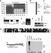
Molecular and biochemical characterization of lister transcript and LISTERIN protein. (A) Haplotype mapping of the lister interval on chromosome 16. All recombinant mice shown are affected. Black squares labeled “B” denote B6 homozygotes, gray squares labeled “H” denote B6;NOD, H-2k heterozygotes, and unlabeled squares are inferred. All physical positions are relative to the National Center for Biotechnology Information Build 34 mouse genome assembly. (B) The lister gene on the mouse chromosome 16. (C) T → A transversion in lister gene. (D) The mutation results in removal of a splice-donor site in lister and an internal deletion of exon 11, while the original reading frame is preserved. (E) Detection of altered mRNA splicing in lister mutant by RT-PCR. (F) Western blot of LISTERIN expression in mouse spinal cord extracts from terminal-stage lister/lister and +/+ mice. (G) Alignment of mouse LISTERIN RING domain and its homologs. The asterisk highlights the conserved zinc-chelating residues in the RING domain. At, Arabidopsis thaliana; Ce, C. elegance; Dm, Drosophila melanogaster; Hs, Human sapien; Mm, Mus musculus. (H) LISTERIN primary structure with a conserved RING finger at its C terminus and the location of the deleted 14 aa in the lister allele. (I) Products of in vitro ubiquitylation assay in a drop-out experiment using the LISTERIN RING finger, revealed by Western blot with anti-ubiquitin antibody.

References

    1. Singleton AB, et al. Alpha-Synuclein locus triplication causes Parkinson's disease. Science. 2003;302:841. - PubMed
    1. Spillantini MG, et al. Alpha-synuclein in Lewy bodies. Nature. 1997;388:839–840. - PubMed
    1. Kruger R, et al. Ala30Pro mutation in the gene encoding alpha-synuclein in Parkinson's disease. Nat Genet. 1998;18:106–108. - PubMed
    1. Zarranz JJ, et al. The new mutation, E46K, of alpha-synuclein causes Parkinson and Lewy body dementia. Ann Neurol. 2004;55:164–173. - PubMed
    1. Farrer MJ. Genetics of Parkinson disease: Paradigm shifts and future prospects. Nat Rev Genet. 2006;7:306–318. - PubMed

Publication types

Substances