. 2009 Feb 24;106(8):2601-6.
doi: 10.1073/pnas.0808220106. Epub 2009 Feb 5.

Rational stabilization of enzymes by computational redesign of surface charge-charge interactions

Affiliations

Rational stabilization of enzymes by computational redesign of surface charge-charge interactions

Alexey V Gribenko et al. Proc Natl Acad Sci U S A. .

Abstract

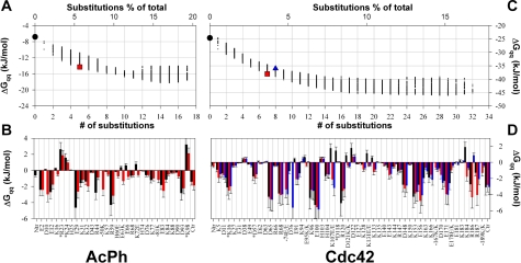
Here, we report the application of a computational approach that allows the rational design of enzymes with enhanced thermostability while retaining full enzymatic activity. The approach is based on the optimization of the energy of charge-charge interactions on the protein surface. We experimentally tested the validity of the approach on 2 human enzymes, acylphosphatase (AcPh) and Cdc42 GTPase, that differ in size (98 vs. 198-aa residues, respectively) and tertiary structure. We show that the designed proteins are significantly more stable than the corresponding WT proteins. The increase in stability is not accompanied by significant changes in structure, oligomerization state, or, most importantly, activity of the designed AcPh or Cdc42. This success of the design methodology suggests that it can be universally applied to other enzymes, on its own or in combination with the other strategies based on redesign of the interactions in the protein core.


Conflict of interest statement

The authors declare no conflict of interest.

Figures

Fig. 1.
Fig. 1.
The diagram structures of 2 proteins studied in this paper. The locations of active sites are shown by van der Waals surfaces for the substrates. Positions of the substitutions are shown by gray circles corresponding to the backbone CA atoms. (A) Acylphosphatase (AcPh): comparison of the structures of bovine protein (PDB code 2ACY) (34) with the 3D structures of human protein AcPh-wt (blue) and its designed variant AcPh-des (green). The structures of AcPh-wt and AcPh-des were determined in this work by using multidimentional NMR spectroscopy (see Materials and Methods and SI Appendix for details). (B) Cdc42: structural model of human protein (PDB code 1AN0) as solved by x-ray crystallography in ref. .
Fig. 2.
Fig. 2.
The dependence of the energy of charge–charge interactions on the number of amino acid substitutions (or the percentage of substitutions relative to the total number of amino acid residues) in AcPh (A) and Cdc42 (C). Each small dot corresponds to a different sequence. The sequences selected for experimental verification are shown in large symbols: AcPh-wt or Cdc42-wt, black circles; AcPh-des, red squares; Cdc42-des1, blue squares; and Cdc42-des2, red triangles. (B and D) The corresponding per residue energies of charge–charge interactions as calculated by TKSA model are given in B (AcPh-wt, black bars; AcPh-des, red bars) and D (Cdc42-wt, black bars; Cdc42-des1, red bars; Cdc42-des2, blue bars). Positive energies represent overall unfavorable interactions of a given residue with all other ionizable residues in the proteins, whereas negative values of ΔGqq reflect overall favorable interactions.
Fig. 3.
Fig. 3.
Biophysical characterization of the designed and WT acylphosphatase (AcPh) and Cdcd42. (A) Comparison of the experimental partial molar heat capacity profiles of AcPh-wt (white circles) and AcPh-des (red squares). Solid lines are the results of the fit according to a 2-state unfolding model. The results of the fit are given in Table 1. (B) Comparison of the experimental temperature-induced unfolding profiles of AcPh-wt (black thin line) and AcPh-des (red thin line) as monitored by the changes in ellipticity at 222 nm. Thick solid lines are the results of the fit according to a 2-state unfolding model. The results of the fit are given in Table 1. (C) Comparison of the experimental temperature induced unfolding profiles of Cdc42-wt (thin black line), Cdc42-des1 (thin blue line), and Cdc42-des2 (thin red line) as monitored by the changes in ellipticity at 222 nm. (D) Dependence of specific activity of Cdc42 variants (Cdc42-wt, black circles; Cdc42-des1, blue squares; Cdc42-des2, red triangles) on temperature. Lines are drawn to guide the eye. (E) Resistance of Cdc42 variants to aggregation after exposure to high temperature. The fraction of soluble monomer remained in solution after exposure to elevated temperatures was measured by using size-exclusion chromatography as described in Materials and Methods. Shown are Cdc42-wt (black circles), Cdc42-des1 (blue squares), and Cdc42-des2 (red triangles), and lines are drawn to guide the eye.
Fig. 4.
Fig. 4.
Stimulation of the GTPase activity of the Cdc42 proteins by the increasing concentrations of Cdc42GAP. Experiments were performed at the initial GTP concentration of 100 μM. Shown are Cdc42-wt (black circles), Cdc42-des1 (blue squares), and Cdc42-des2 (red triangles). Concentration of all 3 Cdc42 proteins was kept constant at 0.5 μM. Data were fitted to a binding equation where y is the fractional saturation, LT is the total Cdc42GAP concentration, MT is the total Cdc42 concentration, and Kd,app is the apparent dissociation constant of Cdc42 to Cdc42GAP. Dashed lines are the results of the fit for each individual protein, and the solid line is the fit of all datapoints to a single Kd,app. See the results of the fit in Table 2.

References

    1. Sanchez-Ruiz JM, Makhatadze GI. To charge or not to charge? Trends Biotechnol. 2001;19:132–135. - PubMed
    1. Dantas G, Kuhlman B, Callender D, Wong M, Baker D. A large scale test of computational protein design: Folding and stability of nine completely redesigned globular proteins. J Mol Biol. 2003;332:449–460. - PubMed
    1. Korkegian A, Black ME, Baker D, Stoddard BL. Computational thermostabilization of an enzyme. Science. 2005;308:857–860. - PMC - PubMed
    1. Malakauskas SM, Mayo SL. Design, structure and stability of a hyperthermophilic protein variant. Nat Struct Biol. 1998;5:470–475. - PubMed
    1. Pokala N, Handel TM. Review: Protein design–where we were, where we are, where we're going. J Struct Biol. 2001;134:269–281. - PubMed

Publication types

LinkOut - more resources