

Analysis of rare APC variants at the mRNA level: six pathogenic mutations and literature review
- PMID: 19196998
- PMCID: PMC2665862
- DOI: 10.2353/jmoldx.2009.080129
Analysis of rare APC variants at the mRNA level: six pathogenic mutations and literature review
Abstract
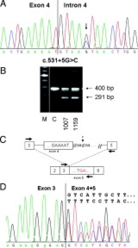
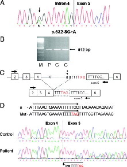
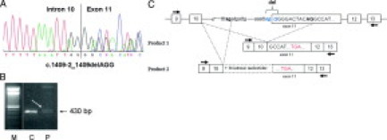
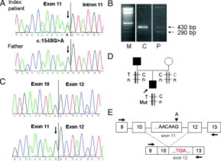
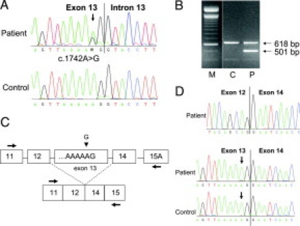
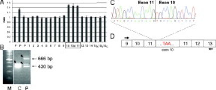
In monogenic disorders, the functional evaluation of rare, unclassified variants helps to assess their pathogenic relevance and can improve differential diagnosis and predictive testing. We characterized six rare APC variants in patients with familial adenomatous polyposis at the mRNA level. APC variants c.531 + 5G>C and c.532-8G>A in intron 4, c.1409-2_1409delAGG in intron 10, c.1548G>A in exon 11, and a large duplication of exons 10 and 11 result in a premature stop codon attributable to aberrant transcripts whereas the variant c.1742A>G leads to the in-frame deletion of exon 13 and results in the removal of a functional motif. Mutation c.1548G>A was detected in the index patient but not in his affected father, suggesting mutational mosaicism. A literature review shows that most of the rare APC variants detected by routine diagnostics and further analyzed at the transcript level were evaluated as pathogenic. The majority of rare APC variants, particularly those located close to exon-intron boundaries, could be classified as pathogenic because of aberrant splicing. Our study shows that the characterization of rare variants at the mRNA level is crucial for the evaluation of pathogenicity and underlying mutational mechanisms, and could lead to better treatment modalities.
Figures
Similar articles
-
Familial adenomatous polyposis: aberrant splicing due to missense or silent mutations in the APC gene.Hum Mutat. 2004 Nov;24(5):370-80. doi: 10.1002/humu.20087. Hum Mutat. 2004. PMID: 15459959
-
Alternative splicing and nonsense-mediated mRNA decay in the regulation of a new adenomatous polyposis coli transcript.Gene. 2007 Jun 15;395(1-2):8-14. doi: 10.1016/j.gene.2006.10.027. Epub 2007 Jan 12. Gene. 2007. PMID: 17360132
-
Transcript dosage effect in familial adenomatous polyposis: model offered by two kindreds with exon 9 APC gene mutations.Hum Mutat. 1998;11(3):197-201. doi: 10.1002/(SICI)1098-1004(1998)11:3<197::AID-HUMU3>3.0.CO;2-F. Hum Mutat. 1998. PMID: 9521420
-
Familial adenomatous polyposis.Best Pract Res Clin Gastroenterol. 2009;23(2):197-207. doi: 10.1016/j.bpg.2009.02.010. Best Pract Res Clin Gastroenterol. 2009. PMID: 19414146 Review.
-
APC germline mutations in individuals being evaluated for familial adenomatous polyposis: a review of the Mayo Clinic experience with 1591 consecutive tests.J Mol Diagn. 2013 Jan;15(1):31-43. doi: 10.1016/j.jmoldx.2012.07.005. Epub 2012 Nov 14. J Mol Diagn. 2013. PMID: 23159591 Review.
Cited by
-
Genotype to phenotype: analyzing the effects of inherited mutations in colorectal cancer families.Mutat Res. 2010 Nov 10;693(1-2):32-45. doi: 10.1016/j.mrfmmm.2009.09.004. Epub 2009 Sep 17. Mutat Res. 2010. PMID: 19766128 Free PMC article. Review.
-
Large intron 14 rearrangement in APC results in splice defect and attenuated FAP.Hum Genet. 2010 Mar;127(3):359-69. doi: 10.1007/s00439-009-0776-9. Epub 2009 Dec 22. Hum Genet. 2010. PMID: 20033212 Free PMC article.
-
Inactivation of promoter 1B of APC causes partial gene silencing: evidence for a significant role of the promoter in regulation and causative of familial adenomatous polyposis.Oncogene. 2011 Dec 15;30(50):4977-89. doi: 10.1038/onc.2011.201. Epub 2011 Jun 6. Oncogene. 2011. PMID: 21643010 Free PMC article. Clinical Trial.
-
Gene-specific ACMG/AMP classification criteria for germline APC variants: Recommendations from the ClinGen InSiGHT Hereditary Colorectal Cancer/Polyposis Variant Curation Expert Panel.Genet Med. 2024 Feb;26(2):100992. doi: 10.1016/j.gim.2023.100992. Epub 2023 Oct 4. Genet Med. 2024. PMID: 37800450 Free PMC article.
-
Allele-specific expression of APC in adenomatous polyposis families.Gastroenterology. 2010 Aug;139(2):439-47, 447.e1. doi: 10.1053/j.gastro.2010.04.047. Epub 2010 Apr 29. Gastroenterology. 2010. PMID: 20434453 Free PMC article.
References
-
- Pagani F, Buratti E, Stuani C, Baralle FE. Missense, nonsense, and neutral mutations define juxtaposed regulatory elements of splicing in cystic fibrosis transmembrane regulator exon 9. J Biol Chem. 2003;278:26580–26588. - PubMed
-
- Pagani F, Stuani C, Tzetis M, Kanavakis E, Efthymiadou A, Doudounakis S, Casals T, Baralle FE. New type of disease causing mutations: the example of the composite exonic regulatory elements of splicing in CFTR exon 12. Hum Mol Genet. 2003;12:1111–1120. - PubMed
-
- Pagani F, Stuani C, Zuccato E, Kornblihtt AR, Baralle FE. Promoter architecture modulates CFTR exon 9 skipping. J Biol Chem. 2003;278:1511–1517. - PubMed
Publication types
MeSH terms
Substances
LinkOut - more resources
Full Text Sources
