Review
. 2009 Mar;174(3):715-21.
doi: 10.2353/ajpath.2009.080758. Epub 2009 Feb 5.

Wnt signaling, lgr5, and stem cells in the intestine and skin

Affiliations
Review

Wnt signaling, lgr5, and stem cells in the intestine and skin

Andrea Haegebarth et al. Am J Pathol. 2009 Mar.

Abstract

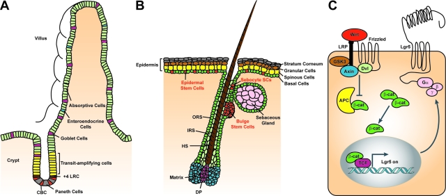
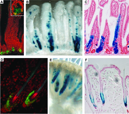
Stem cells hold great promise for regenerative medicine, but have remained elusive in many tissues because of a lack of adequate definitive markers. Progress in mouse genetics has provided the tools for characterization and validation of stem cell markers by functional and/or lineage tracing assays. The Wnt target gene Lgr5 has been recently identified as a novel stem cell marker of the intestinal epithelium and the hair follicle. In the intestine, Lgr5 is exclusively expressed in cycling crypt base columnar cells. Genetic lineage-tracing experiments revealed that crypt base columnar cells are capable of self-renewal and multipotency, thus representing genuine intestinal stem cells. In the stem cell niche of the murine hair follicle, Lgr5 is expressed in actively cycling cells. Transplantation and lineage tracing experiments have demonstrated that these Lgr5(+ve) cells maintain all cell lineages of the hair follicle throughout long periods of time and can build entire new hair follicles. Expression of Lgr5 in multiple other organs indicates that it may represent a global marker of adult stem cells. This review attempts to provide a comprehensive overview of the stem cell compartments in the intestine and skin with a focus on the cycling, yet long-lived and multipotent, Lgr5(+ve) stem cell populations.


Figures

Figure 1
Figure 1
Epithelial SC compartments in the mammalian intestine and the skin and Wnt signaling in SCs. A: The intestinal crypt-villus unit. Intestinal epithelial SCs are thought to reside at the base of the crypt, either in the +4 position counting from the bottom of the crypt (brown) directly above the Paneth cells (gray), or as CBC cells (red) located between the Paneth cells. Recent evidence demonstrates that Lgr5 marks these rapidly cycling CBC cells, which can both self-renew and give rise to TA progenitor cells (yellow). The intestinal TA cells differentiate into four lineages—long-lived Paneth cells (gray) that occupy the base of the crypt and the enteroendocrine cells (blue), goblet cells (purple). and enterocytes (green) that move upwards and cover the villus. B: The skin. Three distinct niches for multipotent epidermal SCs exist—the bulge region of the HF, the base of the SG, and the basal layer of the IFE. SCs of these niches (red) are essential for the self-renewal of their pertinent tissue during normal homeostasis. The IFE SCs give rise to rapidly cycling keratinocytes that differentiate while moving upward to form the spinous layer, granular layer, and stratum corneum. Sebocyte SCs reside near the base of the SG. They give rise to proliferative progeny that differentiates into lipid-filled sebocytes. SCs of the HF are located within the bulge area. After the first and all subsequent hair cycles, bulge cells generate cells of the outer root sheath (ORS), which is thought to contain SCs that continue to migrate down to the follicle base and fuel the proliferative compartment of TA cells located at the base of the hair (matrix cells, blue) and adjacent to the dermal papilla (DP, purple). Matrix cells differentiate to form the various layers of the inner root sheath (IRS) and the hair shaft (HS). Lgr5+ve rapidly cycling cells reside in the bulge and the ORS of the growing HF and give rise to all of the newly formed cells of HF. C: Canonical Wnt signaling in SCs. SCs in the intestine and the skin are regulated by Wnt signaling. Wnt ligands bind to the Frizzled/LRP co-receptor complex and activate the canonical signaling pathway. Axin is recruited to the plasma membrane resulting in the inactivation of the APC destruction complex and subsequent stabilization of β-catenin. β-catenin translocates to the nucleus, where it binds Tcf transcription factors, thus activating the Wnt target gene Lgr5. Lgr5 encodes a seven-transmembrane protein with a large extracellular domain for ligand binding and a short cytoplasmic tail for coupling to G proteins.
Figure 2
Figure 2
Lgr5 expression in the small intestine and the HF. A: Lgr5 expression is restricted to CBC cells interspersed between the Paneth cells at the base of the crypt (inset) of the small intestine of Lgr5-GFP knockin mice. Magnification = ×20 (inset ×60). B and C: Lineage tracing in Lgr5-GFP-Ires-CreERT2/Rosa26-LacZ mice induced for 5 days with low-dose tamoxifen. Whole-mount photograph (B) and section (C) showing a ribbon of blue cells extending from a single CBC cell at the base of the crypt to the tip of the villus. This demonstrates that Lgr5+ve CBC cells are capable of generating the entire villus epithelium. All epithelial cell types are present within the blue ribbons, proving that the Lgr5+ve CBC cells are multipotent. Similar observations at much later time points (6 to 14 months after induction) prove that Lgr5+ve CBC cells are long-lived, allowing the conclusion that they are the true intestinal SC. Magnification = ×16 (B); ×10 (C). D: The Lgr5 gene is exclusively expressed in the lower bulge and the secondary germ of telogen HFs of Lgr5-GFP knockin mice, whereas in anagen and catagen HF Lgr5+ve cells occur in the lower ORS (not shown). Magnification = ×60. E and F: Whole-mount photograph (E) and section (F) from Lgr5-GFP-Ires-CreERT2/Rosa26-LacZ mice 6 months after induction with low-dose tamoxifen at postnatal day 20 (when all HFs are in telogen). All blue cells necessarily originate from Lgr5+ve telogen HF cells. Blue cells occur in all parts of the newly formed HF below the SG, proving that Lgr5+ve HF cells are multipotent as well as long-lived. (Adapted from Barker et al.5) Magnification = ×16 (E); ×20 (F).

Similar articles

Cited by

References

    1. Kiel MJ, Yilmaz OH, Iwashita T, Yilmaz OH, Terhorst C, Morrison SJ. SLAM family receptors distinguish hematopoietic stem and progenitor cells and reveal endothelial niches for stem cells. Cell. 2005;121:1109–1121. - PubMed
    1. Shackleton M, Vaillant F, Simpson KJ, Stingl J, Smyth GK, Asselin-Labat ML, Wu L, Lindeman GJ, Visvader JE. Generation of a functional mammary gland from a single stem cell. Nature. 2006;439:84–88. - PubMed
    1. Stingl J, Eirew P, Ricketson I, Shackleton M, Vaillant F, Choi D, Li HI, Eaves CJ. Purification and unique properties of mammary epithelial stem cells. Nature. 2006;439:993–997. - PubMed
    1. Cairns J. Mutation selection and the natural history of cancer. Nature. 1975;255:197–200. - PubMed
    1. Barker N, van Es JH, Kuipers J, Kujala P, van den Born M, Cozijnsen M, Haegebarth A, Korving J, Begthel H, Peters PJ, Clevers H. Identification of stem cells in small intestine and colon by marker gene Lgr5. Nature. 2007;449:1003–1007. - PubMed

Publication types