. 2009 Mar;174(3):1084-96.
doi: 10.2353/ajpath.2009.080625. Epub 2009 Feb 5.

Chloride intracellular channel protein-4 functions in angiogenesis by supporting acidification of vacuoles along the intracellular tubulogenic pathway

Affiliations

Chloride intracellular channel protein-4 functions in angiogenesis by supporting acidification of vacuoles along the intracellular tubulogenic pathway

Barbara Ulmasov et al. Am J Pathol. 2009 Mar.

Abstract

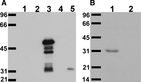
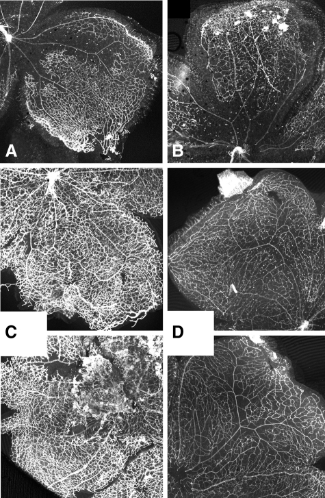
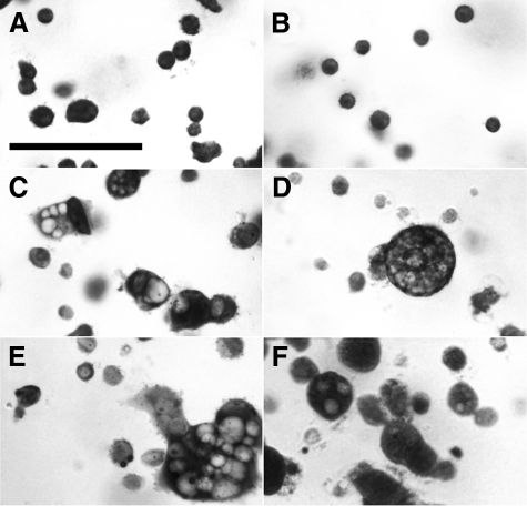
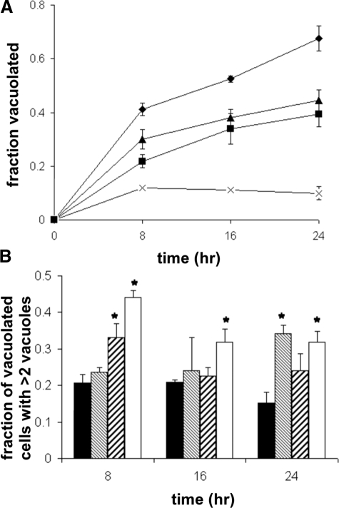
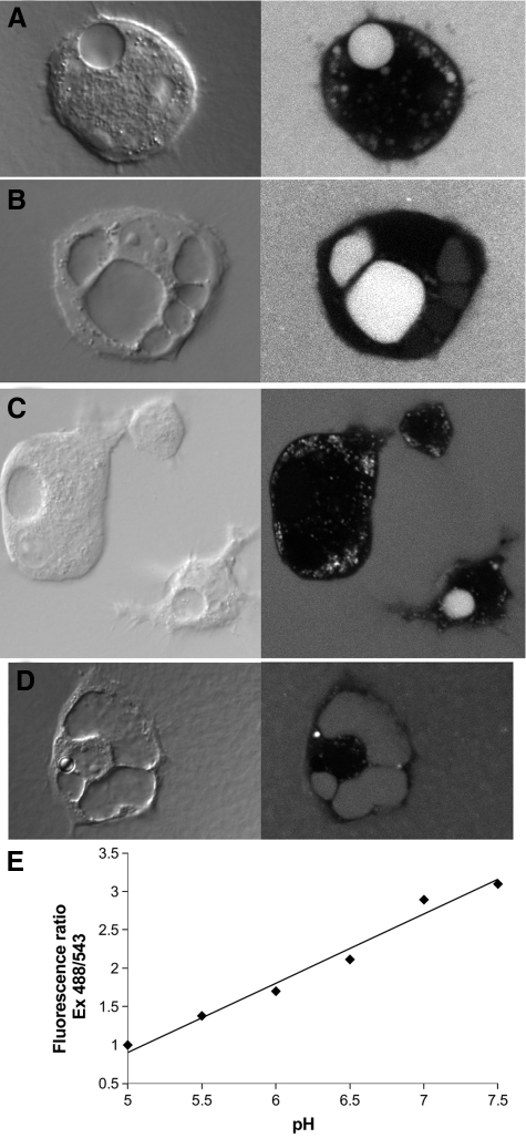
Endothelial cells form capillary tubes through the process of intracellular tubulogenesis. Chloride intracellular channel (CLIC) family proteins have been previously implicated in intracellular tubulogenesis, but their specific role has not been defined. In this study, we show that disruption of the Clic4 gene in mice results in defective angiogenesis in vivo as reflected in a Matrigel plug angiogenesis assay. An angiogenesis defect is also apparent in the retina, both in the decreased spontaneous development of retinal vasculature of unstressed mice and in the dramatically decreased angiogenic response of retinal vessels to an oxygen toxicity challenge. We found that endothelial cells derived from Clic4(-/-) mice demonstrated impaired tubulogenesis in three-dimensional fibrin gels compared with cells derived from wild-type mice. Furthermore, we found that tubulogenesis of wild-type cells in culture was inhibited by both an inhibitor of CLICs and an inhibitor of the vacuolar proton ATPase. Finally, we showed that vacuoles along the endothelial tubulogenesis pathway are acidic in wild-type cells, and that vacuolar acidification is impaired in Clic4(-/-) cells while lysosomal acidification is intact. We conclude that CLIC4 plays a critical role in angiogenesis by supporting acidification of vacuoles along the cell-hollowing tubulogenic pathway.


Figures

Figure 1
Figure 1
Characterization of AP255 anti-CLIC4 antibody. A: Lysates of bacteria expressing glutathione S-transferase (GST, lane 1) or GST-CLIC1 (lane 2), GST-CLIC4 (lane 3), or GST-CLIC5 (lane 4) fusion protein, or 20 μg of mouse kidney homogenate (lane 5) were separated on 10% SDS-PAGE gel, blotted, and probed with a 1:1000 dilution of affinity-purified antibody 255 (AP255). B: Twenty μg of mouse kidney homogenate were separated on 12% SDS-PAGE gel, blotted, and probed with a 1:1000 dilution of AP255 either without (lane 1) or with (lane 2) preabsorption of diluted antibody with the immune peptide at 20 μg/ml for 1 hour prior. Migration positions of molecular weight standards are labeled in kDa. The GST-CLIC4 fusion protein runs at ∼50 kDa (with degradation products below); native mouse CLIC4 runs at 31 kDa.
Figure 2
Figure 2
Targeted disruption of the Clic4 gene. A: Structure of the Clic4 gene (top), the targeting construct (middle), and the structure of the disrupted Clic4 gene after homologous recombination (bottom). N, NheI restriction sites; numbered solid boxes, exons; Neo, neomycin resistance cassette; HSV-TK, herpes simplex virus thymidine kinase gene; Cre, cre recombinase gene under control of tACE promoter. The position of the external probe for Southern blotting is indicated. The horizontal lines show the positions of the 6.9- and 9.6-kb restriction fragments diagnostic for wild-type and properly targeted allele, respectively. B: Southern blot analysis of NheI-digested genomic DNA from wild-type ES cells and a recombinant ES line. The blot was hybridized with 5′ external probe. The wild-type and mutant alleles are indicated by 6.9- and 9.6-kb NheI fragments, respectively. C: Tail clip DNA was prepared from members of a litter and used to direct PCR using the genotyping primers. Sizes of the two major amplification products are indicated in bp. The upper band is the product from the wild-type gene, the lower band is the product from the recombinant gene. Resulting CLIC4 genotypes are lane 1, +/−; lane 2, −/−; lane 3, −/−; lane 4, +/+; lane 5, +/−; lane 6, +/+. D: Fifty μg of total protein from liver or kidney of CLIC4−/− and wild-type mice was separated by SDS-PAGE and probed with antibody AP1058, specific for CLIC4. Lane 1: CLIC4−/− kidney; lane 2: CLIC4−/− liver; lane 3: wild-type kidney; lane 4: wild-type liver. Migration position of the 31-kDa molecular weight standard is indicated by the arrow.
Figure 3
Figure 3
Aberrant Matrigel plug angiogenesis in Clic4−/− mice. A–F: Trichrome-stained sections of Matrigel plugs imaged with ×10 (left) or ×40 (center and right) objective. A–C: Plugs from wild type. D–F: Plugs from Clic4−/−. Note RBC-filled capillaries in B, well-formed capillary lumen in C, and dilated, vesicle-filled cells in E and F. G–K: Sections of Matrigel plugs stained with the endothelial marker ILB4 (red) and the nuclear marker Sytox Green (green). G–H: Plugs from wild-type mice. I–K: Plugs from Clic4−/− mice. Note the lectin-stained highly vacuolated cells in J and K. Scale bars: 100 μm (A, B, D, E); 50 μm (G); 20 μm (K).
Figure 4
Figure 4
CLIC4 is expressed in retinal blood vessels. Mouse retinas were double stained with AP255, specific for CLIC4 (red) and with antibody to the endothelial marker CD31 (green), and images were collected with confocal fluorescence microscopy. A: Whole mount of wild-type retina. There is clear co-localization to retinal surface vessels. B and C: Frozen sections were prepared from whole eyes and stained as above. The dotted line in the green channel image denotes the inner surface of the retina. The rich vascular plexus immediately below the pigmented epithelium is visible in the bottom left corner of each image. B: Wild-type retina. Cross sections of vessels are marked with arrowheads. C: Clic4−/− retina. Scale bars: 75 μm (A); 50 μm (B).
Figure 5
Figure 5
Retinal vasculature of 4-day-old mice. Representative images of retinas from 4-day-old wild-type (left) and Clic4−/− (right) mice stained with ILB4. A and B: Composite low-power images of wild-type (A) and Clic4−/− (B) retinas. C and D: High-power images from the leading edge of the vascular plexus from wild-type (C) and Clic4−/− (D) mice. Tip cells (arrows) tend to show more prominent vacuolization in Clic4−/−. E–H: Large multivacuolated lectin-stained cells (arrows) are visible immediately behind the leading edge of the growing vascular tree, more prominent in the Clic4−/− (F, H) than wild-type (E, G) eyes. G and H: Images of a single microscope field from wild-type (G) or Clic4−/− (H) mice are shown at two different focal planes, one focused on the vascular plexus (left image of each pair) and one focused ∼10 μm higher (right image of each pair).
Figure 6
Figure 6
Retinal vasculature at 7 and 21 days. Representative composite low-power images of lectin-stained retinal whole mounts. A: Seven-day-old wild type. B: Seven-day-old Clic4−/−. C: Twenty-one-day-old wild type. D: Twenty-one-day-old Clic4−/−.
Figure 7
Figure 7
Retinal vascular response to oxygen toxicity. Representative composite low-power images of lectin-stained retinal whole mounts. A and B: Twelve-day-old wild-type (A) and Clic4−/− (B) after 5 days of high (75%) oxygen exposure. C and D: Seventeen-day-old wild-type (C) and Clic4−/− (D) after 5 days of 75% oxygen exposure (upper) and subsequent 5 days of recovery in room air (lower).
Figure 8
Figure 8
Vacuolization of endothelial cells in fibrin gels. Primary cultures of mouse heart endothelial cells were induced to undergo tubulogenesis in fibrin gels and stained with toluidine blue. A and B: Wild-type (A) and Clic4−/− (B) cells immediately after plating. Note homogenous populations of small round nonvacuolated cells in both. C: Wild-type cells after 16 hours of culture. Small nonvacuolated cells and cells with a single large vacuole or multiple vacuoles are all present. D: Clic4−/− cells after 16 hours of culture. Fewer cells are vacuolated and multivacuolated cells are more prominent. E: Wild-type cells after 16 hours in the presence of 200 μmol/L IAA-94. F: Wild-type cells after 16 hours in the presence of 100 μmol/L bafilomycin. Scale bar = 50 μm.
Figure 9
Figure 9
Time course of endothelial cell vacuolization in culture. Endothelial cells were cultured in fibrin gels, fixed at various time points, stained with toluidine blue, and observed under light microscopy. The fraction of total cells vacuolated and fraction of vacuolated cells containing three or more vacuoles were determined. A: Plot of fraction of cells vacuolated throughout time. Results shown are averages from three independent experiments performed with different cell preparations. Diamonds, wild-type cells; squares, Clic4−/− cells; triangles, wild-type cells grown in the presence of 200 μmol/L IAA-94; crosses, wild-type cells grown in the presence of 100 nmol/L bafilomycin. Error bars represent SE of mean. Error bars smaller than the symbols are not visible. P < 0.05 for comparisons between wild-type and all other groups at all points. B: Fraction of vacuolated cells that contained three or more clearly distinguishable vacuoles at each time point. Values are averaged from three independent experiments. Solid bars, wild-type cells; fine hatched bars, wild-type with IAA-94; coarse hatched bars, wild-type with bafilomycin; open bars, Clic4−/− cells. Error bars represent SEM. *P < 0.05 when compared with wild type. Significance was determined using analysis of variance.
Figure 10
Figure 10
Immunolocalization of CLIC4 in endothelial cells undergoing vacuolization in fibrin gels. Cells were cultured in fibrin gels for 16 hours, fixed, and stained with AP255 antibody to CLIC4. Confocal fluorescent and matching DIC images were obtained. A: Wild-type cells. B: Clic4−/− cells. C: Wild-type cells cultured in the presence of 100 nmol/L bafilomycin. Scale bar = 10 μm.
Figure 11
Figure 11
Loading of endothelial vacuoles with pH-sensitive dextran. Cells were grown in fibrin gels supplemented with fluorescent dextran for 8 to 12 hours. A–C: Paired DIC (left) and confocal fluorescence (right) images of wild-type cells are shown. Large vacuoles are obvious in the DIC images and show variable loading with dextran. Abundant, much smaller endosomal/lysosomal structures are also apparent. D: Clic4−/− cell loaded with dextran and imaged as above. E: Relationship between pH and fluorescence ratio from one fibrin gel. All panels are printed at the same magnification. The vertical dimension of panel A is 50 microns.

Similar articles

Cited by

References

    1. Lubarsky B, Krasnow MA. Tube morphogenesis: making and shaping biological tubes. Cell. 2003;112:19–28. - PubMed
    1. Kamei M, Saunders WB, Bayless KJ, Dye L, Davis GE, Weinstein BM. Endothelial tubes assemble from intracellular vacuoles in vivo. Nature. 2006;442:453–456. - PubMed
    1. Béraud-Dufour S, Balch W. A journey through the exocytic pathway. J Cell Sci. 2002;115:1779–1780. - PubMed
    1. Mellman I. The importance of being acid: the role of acidification in intracellular membrane traffic. J Exp Biol. 1992;172:39–45. - PubMed
    1. Ashley RH. Challenging accepted ion channel biology: p64 and the CLIC family of putative intracellular anion channel proteins. Mol Membr Biol. 2003;20:1–11. - PubMed

Publication types