. 2009 Mar 27;104(6):733-41.
doi: 10.1161/CIRCRESAHA.108.183053. Epub 2009 Feb 5.

Smooth muscle cells give rise to osteochondrogenic precursors and chondrocytes in calcifying arteries

Affiliations

Smooth muscle cells give rise to osteochondrogenic precursors and chondrocytes in calcifying arteries

Mei Y Speer et al. Circ Res. .

Abstract

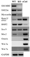
Vascular calcification is a major risk factor for cardiovascular morbidity and mortality. To develop appropriate prevention and/or therapeutic strategies for vascular calcification, it is important to understand the origins of the cells that participate in this process. In this report, we used the SM22-Cre recombinase and Rosa26-LacZ alleles to genetically trace cells derived from smooth muscle. We found that smooth muscle cells (SMCs) gave rise to osteochondrogenic precursor- and chondrocyte-like cells in calcified blood vessels of matrix Gla protein deficient (MGP(-/-)) mice. This lineage reprogramming of SMCs occurred before calcium deposition and was associated with an early onset of Runx2/Cbfa1 expression and the downregulation of myocardin and Msx2. There was no change in the constitutive expression of Sox9 or bone morphogenetic protein 2. Osterix, Wnt3a, and Wnt7a mRNAs were not detected in either calcified MGP(-/-) or noncalcified wild-type (MGP(+/+)) vessels. Finally, mechanistic studies in vitro suggest that Erk signaling might be required for SMC transdifferentiation under calcifying conditions. These results provide strong support for the hypothesis that adult SMCs can transdifferentiate and that SMC transdifferentiation is an important process driving vascular calcification and the appearance of skeletal elements in calcified vascular lesions.


Figures

Figure 1
Figure 1. Genetic tracing and characterization of cells that transcribe β-galactosidase transgene
A. Schematic description of Cre recombination that leads to LacZ gene activation in cells expressing SM22α. B – K. X-gal stained tissues dissected from MGP+/+ (B – H) and MGP−/− (I – K) mice that carry R26R–LacZ and SM22-Cre transgenes (C – J) or R26R–LacZ transgene only (B, K). B, C, and I – K, aorta, D, spleen, E, liver, F, trachea, G, growth plate of a femur, H, marrow of a femur. Sections were counterstained by nuclear fast red (B – H, K) or haematoxylin (J). MGP−/− aorta was stained for mineral by von Kossa method (I). L=lumen; M=media; Ad=adventitia.
Figure 2
Figure 2. Transdifferentiation of SMCs in calcified arteries of MGP−/− mice
Aortas were dissected from 4-week-old SM22α-Cre+/0:R26R+/0:MGP+/+ (AD) and SM22α-Cre+/0:R26R+/0:MGP−/− (EH) mice. Cells of SMC origin were stained by X-gal before embedding. Adjacent sections were stained for mineral by von Kossa method (A and E) and for various cell markers by immunohistochemistry: B and F, SMMHC, C and G, SM22α, D and H, osteopontin. Insert: higher power magnification of the boxed region shows co-localization of β-galactosidase (blue) and osteochondrogenic marker, osteopontin (brown). L =lumen; M=media; Ad=adventitia.
Figure 3
Figure 3. SMCs gave rise to chondrocyte-like cells in calcifying MGP−/− vessels
Aortas were dissected from 6-week-old SM22α-Cre+/0:R26R+/0:MGP−/− mice. Cells of SMC origin were stained by X-gal before embedding. Adjacent sections were stained by haematoxylin and eosin (A), Alizarin red S (B), osteopontin (C), and type II collagen (D). Arrows designate chondrocytes. Insert: higher power magnification of the boxed region shows co-localization of β-galactosidase (blue) and osteochondrogenic marker, osteopontin (brown) and chondrocyte marker, type II collagen (brown). L =lumen; M=media; Ad=adventitia.
Figure 4
Figure 4. Expression of genes associated with differentiation of SMCs, osteoblasts, and chondrocytes in mouse arteries
Total RNA was extracted from 2 – 4 carotids of 2-week-old MGP−/− or MGP+/+ mice. One µg total RNA of these pooled samples was reverse transcribed to cDNA. Various gene expression levels were determined by RT-PCR using specific primers as listed in Online Table 1. One µg total RNA extracted from cementoblasts was used as positive controls for osteochondrogenic genes. Expression levels of a housekeeping gene, glyceraldehyde 3-phosphate dehydrogenase (GAPDH), were determined to assure equal loading. Data shown is one of the two preparations; similar results were achieved in another independent preparation.
Figure 5
Figure 5. Runx2/Cbfa1 expression in MGP−/− arteries of various ages
Aortas were dissected from 1-week-old (H), 2-week-old (AD), 4-week-old (E and F), and 6-week-old (G) SM22α-Cre+/0:R26R+/0:MGP−/− mice. Adjacent sections were stained immunohistochemically for Runx2/Cbfa1 (A, FH), β-galatosidase (B), and SM22α (C), and for mineral by von Kossa (D and E). B and C were counterstained with nuclear fast red, D and E were counterstained with Methyl green, F and G were pre-stained with X-gal. Arrows designate Runx2/Cbfa1 positive cells that originate from SM lineage (β-galactosidase positive). Insert: higher power magnification of the boxed region shows co-localization of β-galactosidase (blue) and Runx2/Cbfa1 (brown). L =lumen; M=media; Ad=adventitia.
Figure 6
Figure 6. SMC calcification and transdifferentiation in culture upon exposure to elevated inorganic phosphate
SMCs were cultured in DMEM culture medium containing basal (1.0 mmol/L) or high (3.0 mmol/L) inorganic phosphate levels. At day 5 calcium content of the cultures was determined as described in “Methods”. Data shown are mean ± SD, n = 3 (A). SMCs treated as in (A) were double stained by X-gal and antibodies specific for SMC lineage proteins (B) and bone tissue associated proteins (C). Similar results were achieved in another independent experiment. ALP=alkaline phosphatase.
Figure 7
Figure 7. Erk signaling in SMC transdifferentiation to osteochondrogenic precursors
SMCs were cultured in the presence (+) or absence (−) of 3.0 mmol/L inorganic phosphate for various days (A), or in the presence of 3.0 mmol/L inorganic phosphate with or without MEK inhibitor U-0126 for 4 days (BD). Cell lysate was collected for Western blot analysis (A and B), and total RNA was extracted for real-time RT-PCR (C and D). Similar results were obtained in another independent experiment.
Figure 8
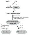
Figure 8. Proposed mechanisms of SMC transdifferentiation in calcified arteries
Loss of MGP with subsequent release from inhibition of BMP2, or exposure to elevated phosphate, leads to increased BMP2 and phosphorylation of Erk1/2 in SMCs. Erk1/2 activation increases Runx2/Cbfa1 and decreases myocardin and SMC lineage markers to generate the osteochondrogenic precursor state. In the presence of high Sox9, and the absence of Msx2, Wnts and osterix, osteochondrogenic precursors preferentially differentiate towards a chondrocytic lineage.

Comment in

References

    1. Rutsch F, Ruf N, Vaingankar S, Toliat MR, Suk A, Hohne W, Schauer G, Lehmann M, Roscioli T, Schnabel D, Epplen JT, Knisely A, Superti-Furga A, McGill J, Filippone M, Sinaiko AR, Vallance H, Hinrichs B, Smith W, Ferre M, Terkeltaub R, Nurnberg P. Mutations in ENPP1 are associated with 'idiopathic' infantile arterial calcification. Nat Genet. 2003;34:379–381. - PubMed
    1. Block GA. Control of serum phosphorus: implications for coronary artery calcification and calcific uremic arteriolopathy (calciphylaxis) Curr Opin Nephrol Hypertens. 2001;10:741–747. - PubMed
    1. Taylor AJ, Burke AP, O'Malley PG, Farb A, Malcom GT, Smialek J, Virmani R. A comparison of the Framingham risk index, coronary artery calcification, and culprit plaque morphology in sudden cardiac death. Circulation. 2000;101:1243–1248. - PubMed
    1. Shanahan CM, Cary NR, Salisbury JR, Proudfoot D, Weissberg PL, Edmonds ME. Medial localization of mineralization-regulating proteins in association with Monckeberg’s sclerosis: evidence for smooth muscle cell-mediated vascular calcification. Circulation. 1999;100:2168–2176. - PubMed
    1. Moe SM, O'Neill KD, Duan D, Ahmed S, Chen NX, Leapman SB, Fineberg N, Kopecky K. Medial artery calcification in ESRD patients is associated with deposition of bone matrix proteins. Kidney Int. 2002;61:638–647. - PubMed

Publication types

MeSH terms

Substances