. 2009 Mar;119(3):512-23.
doi: 10.1172/JCI35070. Epub 2009 Feb 9.

Endothelial-derived FGF2 contributes to the progression of pulmonary hypertension in humans and rodents

Affiliations

Endothelial-derived FGF2 contributes to the progression of pulmonary hypertension in humans and rodents

Mohamed Izikki et al. J Clin Invest. 2009 Mar.

Abstract

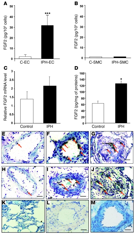
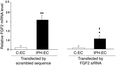
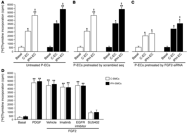
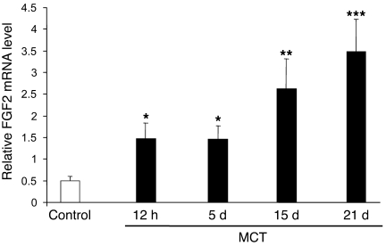
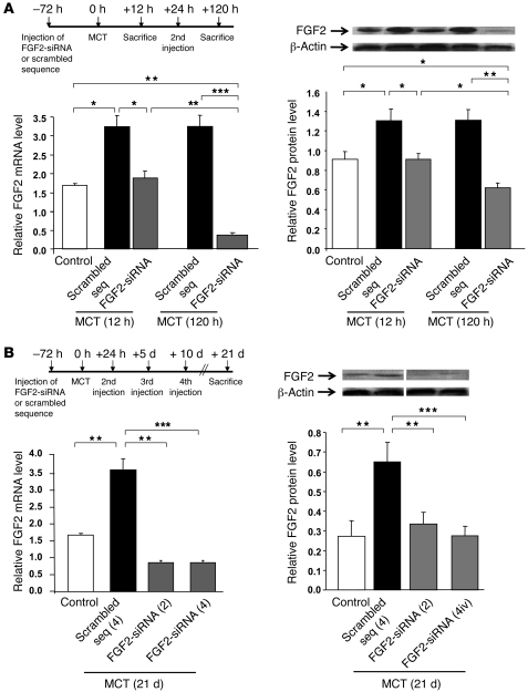
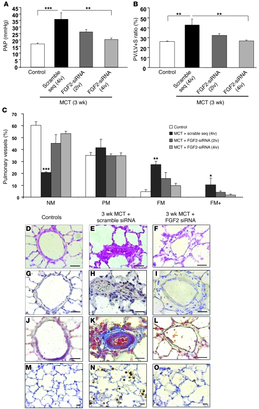
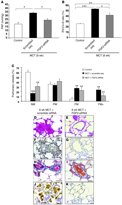
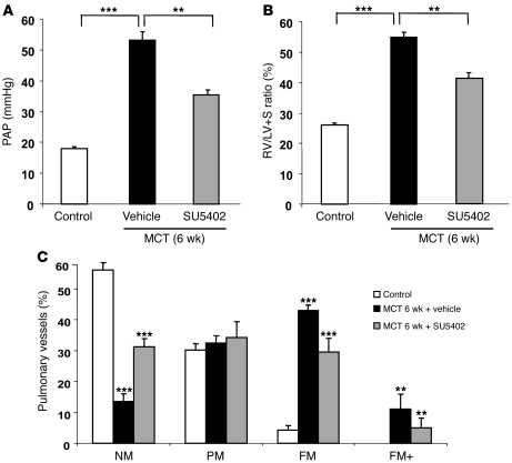
Pulmonary hypertension (PH) is a progressive, lethal lung disease characterized by pulmonary artery SMC (PA-SMC) hyperplasia leading to right-sided heart failure. Molecular events originating in pulmonary ECs (P-ECs) may contribute to the PA-SMC hyperplasia in PH. Thus, we exposed cultured human PA-SMC to medium conditioned by P-EC from patients with idiopathic PH (IPH) or controls and found that IPH P-EC-conditioned medium increased PA-SMC proliferation more than control P-EC medium. Levels of FGF2 were increased in the medium of IPH P-ECs over controls, while there was no detectable difference in TGF-beta1, PDGF-BB, or EGF levels. No difference in FGF2-induced proliferation or FGF receptor type 1 (FGFR1) mRNA levels was detected between IPH and control PA-SMCs. Knockdown of FGF2 in P-EC using siRNA reduced the PA-SMC growth-stimulating effects of IPH P-EC medium by 60% and control P-EC medium by 10%. In situ hybridization showed FGF2 overproduction predominantly in the remodeled vascular endothelium of lungs from patients with IPH. Repeated intravenous FGF2-siRNA administration abolished lung FGF2 production, both preventing and nearly reversing a rat model of PH. Similarly, pharmacological FGFR1 inhibition with SU5402 reversed established PH in the same model. Thus, endothelial FGF2 is overproduced in IPH and contributes to SMC hyperplasia in IPH, identifying FGF2 as a promising target for new treatments against PH.


Figures

Figure 1
Figure 1. FGF2 expression in cultured P-ECs, PA-SMCs, and lungs from patients with IPH.
(A) FGF2 expression by P-ECs and (B) PA-SMCs from controls (C-EC and C-SMC, respectively) and from patients with IPH (IPH-EC and IPH-SMC, respectively). Values are mean ± SEM from 5 controls and 5 patients with IPH. (C) FGF2 mRNA levels and (D) FGF2 protein levels in lung tissues from controls and patients with IPH. Values are mean ± SEM from 12 controls and 14 patients for mRNA levels and 5 controls and 5 patients for protein levels. (EM) In situ hybridization in human pulmonary arteries from controls (E and H) and from patients with IPH (F, G, I, J) using 3′-[35S]–labeled oligonucleotide probes specific for human FGF2 or using a sense probe (KM). Arrows indicate the localization of FGF2 mRNA in the vascular endothelium of muscular pulmonary arteries. *P < 0.05; ***P < 0.001 vs. P-ECs, PA-SMCs, or lungs from control subjects. Scale bars: 100 μm.
Figure 2
Figure 2. FGF2 mRNA level in P-ECs assessed by real-time quantitative PCR.
FGF2 mRNA levels assessed by real-time quantitative PCR in P-ECs obtained from lungs of patients with IPH or controls and treated with FGF2-siRNA or the scrambled siRNA sequence. Values are mean ± SEM from 5 controls and 5 patients with IPH. *P < 0.05; **P < 0.001 vs. P-ECs from control subjects; P < 0.01 between P-ECs treated with FGF2-siRNA vs. scrambled siRNA sequence.
Figure 3
Figure 3. Effects of FGF2 release from P-ECs on PA-SMC growth.
(A) Proliferation of PA-SMCs from controls and patients with IPH stimulated with medium of untreated P-ECs from controls and patients with IPH, (B) with medium from P-ECs pretreated with the scrambled siRNA sequence (seq), or (C) with medium from P-ECs pretreated with FGF2-siRNA. (D) Proliferation of PA-SMCs from controls and patients in response to PDGF (10 ng/ml) or to FGF2 (10 ng/ml) with or without the PDGFR antagonist imatinib, the EGFR inhibitor, or the FGFR1 antagonist SU5402 (10–5 M each). Values are mean ± SEM of 4 experiments. *P < 0.01; **P < 0.001 vs. basal conditions; P < 0.01 between stimulation with medium of P-ECs from IPH vs. P-ECs from controls.
Figure 4
Figure 4. Lung FGF2 expression in MCT-induced PH.
Kinetics of FGF2 mRNA levels measured by real-time quantitative PCR over a 21-day period in lungs of rats (n = 5 in each group) injected s.c. with MCT (60 mg/kg). *P < 0.05; **P < 0.01; ***P < 0.001 vs. control rats injected with saline instead of MCT.
Figure 5
Figure 5. Validation of acute and chronic lung FGF2 knockdown by siRNA.
(A) FGF2 mRNA and protein levels in rat lung 12 hours and 120 hours after MCT injection. Rats were injected with FGF2-siRNA or the scrambled siRNA sequence either once, 3 days before MCT injection (12-hour group), or twice, 3 days before and 1 day after MCT injection (120-hour group). (B) FGF2 mRNA and protein levels in rat lungs 21 days after MCT injection. Rats were injected with FGF2-siRNA or the scrambled siRNA sequence (2 [2iv] or 4 [4iv] injections). FGF2 mRNA levels were assessed using real-time quantitative PCR and protein levels with FGF2 immunoblotting (the lanes were run on the same gel but were not contiguous). All values are mean ± SEM from at least 5 animals in each group. *P < 0.05; **P < 0.01; ***P < 0.001 vs. rats injected with saline instead of MCT or vs. rats treated with the scrambled siRNA sequence.
Figure 6
Figure 6. Evaluation of the efficacy of FGF2-siRNA pretreatment in MCT-induced PH.
Results obtained in rats treated with FGF2-siRNA or the scrambled siRNA sequence before the MCT injection and studied on day 21. Compared with the scrambled siRNA sequence, FGF2-siRNA significantly prevented the development of PH, as assessed by (A) PAP; (B) RV hypertrophy reflected by the RV/(LV + S) weight ratio; and (C) percentages of NM, PM, FM, and FM+ intra-acinar vessels. (DF) Hematoxylin-phloxine-saffron stain; (GI) PCNA immunostaining; (JL) Masson trichrome stain; and (MO) CD68 immunostaining of rat lungs. *P < 0.05; **P < 0.01; ***P < 0.001 vs. rats injected with saline instead of MCT or vs. rats treated with scrambled siRNA sequence. Scale bars: 25 μm.
Figure 7
Figure 7. Evaluation of the efficacy of FGF2-siRNA treatment in MCT-induced PH.
Results obtained in rats treated with FGF2-siRNA or the scrambled siRNA sequence (2 injections) on day 21 and day 25 after MCT injection (treatment) and studied on day 42. Compared with the scrambled siRNA sequence, FGF2-siRNA significantly reversed the development of PH, as assessed by (A) PAP; (B) RV hypertrophy reflected by the RV/(LV + S) weight ratio; and (C) percentages of NM, PM, FM, and FM+ intra-acinar vessels. (D and E) Hematoxylin-phloxine-saffron stain; (F and G) PCNA immunostaining; (H and I) Masson trichrome stain; and (J and K) CD68 immunostaining of rat lungs. *P < 0.05; **P < 0.01; ***P < 0.001 vs. rats injected with saline instead of MCT or vs. rats treated with scrambled siRNA sequence. Scale bars: 25 μm.
Figure 8
Figure 8. Reversal of MCT-induced PH with the FGFR1 inhibitor SU5402.
Results obtained in rats treated with SU5402 (25 mg × kg–1 × d–1) or vehicle (saline) from day 21 to day 42 after MCT injection (treatment) and studied on day 42. Compared with vehicle, SU5402 significantly reversed the development of PH, as assessed by (A) PAP; (B) RV hypertrophy reflected by the RV/(LV + S) weight ratio; and (C) percentages of NM, PM, FM, and FM+ intra-acinar vessels. **P < 0.01; ***P < 0.001 vs. rats injected with saline instead of MCT vs. rats treated with saline instead of SU5402.

References

    1. Rubin L.J. Primary pulmonary hypertension. N. Engl. J. Med. 1997;336:111–117. doi: 10.1056/NEJM199701093360207. - DOI - PubMed
    1. Humbert M., et al. Cellular and molecular pathobiology of pulmonary arterial hypertension. J. Am. Coll. Cardiol. 2004;43:13S–24S. - PubMed
    1. Eddahibi S., et al. Cross talk between endothelial and smooth muscle cells in pulmonary hypertension: critical role for serotonin-induced smooth muscle hyperplasia. Circulation. 2006;113:1857–1864. doi: 10.1161/CIRCULATIONAHA.105.591321. - DOI - PubMed
    1. Armulik A., Abramsson A., Betsholtz C. Endothelial/pericyte interactions. Circ. Res. 2005;97:512–523. doi: 10.1161/01.RES.0000182903.16652.d7. - DOI - PubMed
    1. Dewachter L., et al. Angiopoietin/Tie2 pathway influences smooth muscle hyperplasia in idiopathic pulmonary hypertension. Am. J. Respir. Crit. Care Med. 2006;174:1025–1033. doi: 10.1164/rccm.200602-304OC. - DOI - PubMed

Publication types

MeSH terms

Substances