. 2009 Mar 18;28(6):652-62.
doi: 10.1038/emboj.2009.15. Epub 2009 Feb 5.

Mislocalization of the MRN complex prevents ATR signaling during adenovirus infection

Affiliations

Mislocalization of the MRN complex prevents ATR signaling during adenovirus infection

Christian T Carson et al. EMBO J. .

Abstract

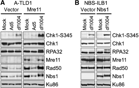
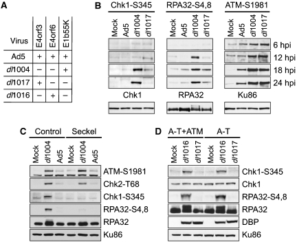
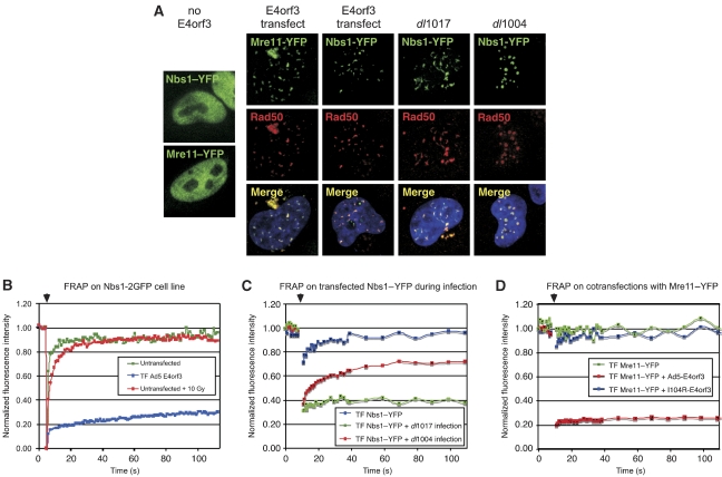
The protein kinases ataxia-telangiectasia mutated (ATM) and ATM-Rad3 related (ATR) are activated in response to DNA damage, genotoxic stress and virus infections. Here we show that during infection with wild-type adenovirus, ATR and its cofactors RPA32, ATRIP and TopBP1 accumulate at viral replication centres, but there is minimal ATR activation. We show that the Mre11/Rad50/Nbs1 (MRN) complex is recruited to viral centres only during infection with adenoviruses lacking the early region E4 and ATR signaling is activated. This suggests a novel requirement for the MRN complex in ATR activation during virus infection, which is independent of Mre11 nuclease activity and recruitment of RPA/ATR/ATRIP/TopBP1. Unlike other damage scenarios, we found that ATM and ATR signaling are not dependent on each other during infection. We identify a region of the viral E4orf3 protein responsible for immobilization of the MRN complex and show that this prevents ATR signaling during adenovirus infection. We propose that immobilization of the MRN damage sensor by E4orf3 protein prevents recognition of viral genomes and blocks detrimental aspects of checkpoint signaling during virus infection.


Figures

Figure 1
Figure 1
The Mre11 and Nbs1 proteins are required for robust ATR signaling in response to mutant Ad infection. (A) Infections were performed in a transformed cell line deficient for Mre11 (A-TLD1) that was transduced with an empty retrovirus vector (vector) or a vector expressing the wild-type Mre11 cDNA (Mre11). Cells were uninfected (Mock) or infected with wild-type Ad5 (Ad5) and the E4-deletion mutant (dl1004). Immunoblotting of lysates prepared at 30 hpi used antibodies for total protein (Chk1, RPA32, Mre11, Rad50 and Nbs1) or phospho-specific sites (Chk1-S345). Ku86 served as a loading control. (B) Infections were performed in the transformed NBS cell line (NBS-ILB1) transduced with an empty retrovirus vector (vector) or a vector expressing the wild-type Nbs1 cDNA.
Figure 2
Figure 2
Phosphorylation of ATR substrates is reduced in the presence of Ad5-E4orf3 and is independent of ATM. (A) Genotypes of mutant viruses used for infection (Bridge and Ketner, 1990). (B) HeLa cells were mock infected or infected with indicated viruses. Cells were harvested at 6, 12, 18 and 24 hpi and prepared for analysis by immunoblotting using antibodies to phospho-specific residues (Chk1-S345, RPA32-S4,8 and ATM-S1981). Below are lysates at 24 hpi probed with antibodies against total protein (Chk1, RPA32 and Ku86) to serve as loading controls. (C) Immunoblots of infections in control cells (48BR) or Seckel cells with mutant ATR (GM18366). (D) Immunoblots of infections in A-T cells with mutant ATM (AT221JE-T) or a complemented line (A-T+ATM). DBP was a marker of virus infection and Ku86 served as a loading control.
Figure 3
Figure 3
Signaling by ATR at viral replication centres. (A) E4orf3 sequesters Nbs1 into tracks away from viral replication centres. HeLa cells were mock infected or infected with indicated viruses. Cells were fixed at 16 hpi, and immunofluorescence was performed. Staining shows that Nbs1 but not RPA32 is excluded from viral replication centres in the presence of E4orf3. (B) Colocalization of the MRN complex with RPA32 at replication centres correlates with RPA32 phosphorylation. Immunofluorescence with a phospho-specific antibody to RPA32-S4,8 shows colocalization with viral replication centres stained with an antibody to DBP only in the absence of E4orf3. (C) Accumulation of RPA32, ATR and ATRIP at viral centres is independent of E4. (D) TopBP1 accumulates at viral centres independently of E4. (E) Activation is observed for ATM, but not ATR, in Seckel cells. (F) ATR signaling is restored in A-TLD cells by expression of Mre11 or the nuclease-defective mutant Mre11-3. In all images, the nuclei were located by co-staining cellular DNA with DAPI (blue).
Figure 4
Figure 4
A region in Ad5-E4orf3 important for targeting the MRN complex. (A) Sequence alignment of E4orf3 proteins. E4orf3 sequences from different human (subgroup noted in brackets) and simian adenoviruses were aligned using the CLUSTAL algorithm and the region around residue I104 is shown. Conserved residues are shown boxed, with CLUSTAL colour scheme reflecting amino acids of similar chemical nature. The I104 residue is highlighted, showing that this site differs between subgroup C and all other sequenced E4orf3 genes. (B) The Ad5 E4orf3-I104R mutant does not redistribute the MRN complex. Plasmids for wild-type and mutant E4orf3 were transfected into HeLa cells. Immunofluorescence shows that the E4orf3 mutant protein still forms tracks and disrupts PML structures but is unable to redistribute members of the MRN complex, which remain diffusely nuclear. Representative images are shown and nuclei are located by co-staining cellular DNA with DAPI.
Figure 5
Figure 5
ATR signaling in response to virus infection is abrogated by E4orf3 proteins that mislocalize the MRN complex. HeLa cells were transfected with an empty plasmid (vector) or with vectors expressing E4orf3 proteins. After 24 h, cells were either mock infected, infected with the E1b55K/E4orf6 mutant (dl1017) (positive control), or infected with the E4-deletion mutant (dl1004). (A, B) Lysates from cells at 24 hpi were immunoblotted with antibodies to Chk1-S345, RPA32-S4,8, ATM-1981 and RPA32. (C) Cells were fixed at 18 hpi, and immunofluorescence was performed with an antibody to RPA32-S4,8. Images are shown merged with DAPI staining. A plasmid expressing GFP was cotransfected with that for E4orf3 at a ratio of 1:10 and GFP staining is shown in the lower left insert panel as a positive control for transfection.
Figure 6
Figure 6
E4orf3 abrogates MRN function by immobilizing the MRN complex. (A) HeLa cells were transfected with Nbs1–YFP or Mre11–YFP alone or together with Ad5-E4orf3 plasmid vector. After 24 h, cells were infected with dl1004 or dl1017. Cells were fixed 18 hpi, and immunofluorescence was performed with a Rad50 antibody. Images shown are merged with DAPI staining. (BD) FRAP analysis of Nbs1 and Mre11 in cells expressing E4orf3 by infection or transfection. The unbleached portion of the cell served to normalize the overall fluorescence decay during the repeated image collection. Arrows above indicate the time of bleaching. (B) Stable U2OS cells expressing Nbs1–2GFP were either untreated, transfected with E4orf3, or treated with 10 Gy gamma-irradiation. (C) HeLa cells were transfected with Nbs1–YFP and then mock treated or infected with dl1004 and dl1017. Cells were analyzed by FRAP at 18 hpi. (D) HeLa cells transfected with Mre11–YFP together with either empty vector or vectors expressing Ad5-E4orf3 and E4orf3-I104R were analyzed by FRAP.
Figure 7
Figure 7
E4orf3 prevents ATR-dependent damage signaling induced by nonviral sources. (A) U2OS cells transfected with a plasmid vector expressing Ad5-E4orf3 were laser microirradiated. Cells were fixed and stained for endogenous Nbs1 and Mdc1. Arrows indicate cells with E4orf3-induced tracks of Nbs1. (B) Stable U2OS cell lines expressing Nbs1–2GFP were transfected with a plasmid expressing Ad5-E4orf3, together with prRFP-C1, which was used as a marker for transfected cells. Cells were laser microirradiated (as indicated by dashed line) and images show the recruitment of Nbs1 to sites of damage. (C) HeLa cells were mock infected, or infected with E1-deleted recombinant Ads expressing GFP (rAd-GFP) or E4orf3 (rAd-E4orf3). At 24 hpi, the cells were mock treated or treated with 2 mM hydroxyurea (HU) for 2 h, and the cells were harvested for immunoblotting. Cellular proteins were detected with antibodies to RPA32 and specific phosphorylated sites at RPA-S4,8 and Nbs1-S343. Ku86 served as a loading control.

References

    1. Adams KE, Medhurst AL, Dart DA, Lakin ND (2006) Recruitment of ATR to sites of ionising radiation-induced DNA damage requires ATM and components of the MRN protein complex. Oncogene 25: 3894–3904 - PMC - PubMed
    1. Araujo FD, Stracker TH, Carson CT, Lee DV, Weitzman MD (2005) Adenovirus type 5 E4orf3 protein targets the Mre11 complex to cytoplasmic aggresomes. J Virol 79: 11382–11391 - PMC - PubMed
    1. Arias EE, Walter JC (2007) Strength in numbers: preventing rereplication via multiple mechanisms in eukaryotic cells. Genes Dev 21: 497–518 - PubMed
    1. Bakkenist CJ, Kastan MB (2003) DNA damage activates ATM through intermolecular autophosphorylation and dimer dissociation. Nature 421: 499–506 - PubMed
    1. Binz SK, Sheehan AM, Wold MS (2004) Replication protein A phosphorylation and the cellular response to DNA damage. DNA Repair 3: 1015–1024 - PubMed

Publication types

MeSH terms