Review
. 2009 Jan;10(1):9-20.
doi: 10.1038/nrm2609.

The 3Ms of central spindle assembly: microtubules, motors and MAPs

Affiliations
Review

The 3Ms of central spindle assembly: microtubules, motors and MAPs

Michael Glotzer. Nat Rev Mol Cell Biol. 2009 Jan.

Abstract

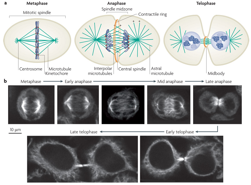
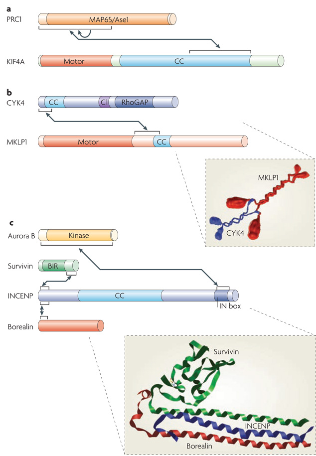
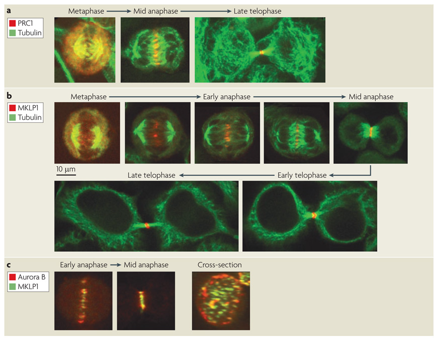
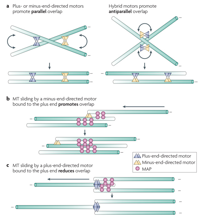
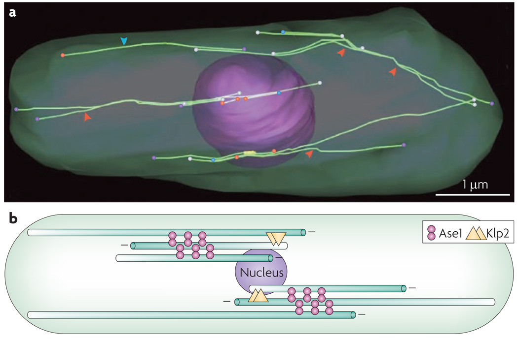
During metaphase, sister chromatids are positioned at the midpoint of the microtubule-based mitotic spindle in preparation for their segregation. The onset of anaphase triggers inactivation of the key mitotic kinase cyclin-dependent kinase 1 (CDK1) and the polewards movement of sister chromatids. During anaphase, the mitotic spindle reorganizes in preparation for cytokinesis. Kinesin motor proteins and microtubule-associated proteins bundle the plus ends of interpolar microtubules and generate the central spindle, which regulates cleavage furrow initiation and the completion of cytokinesis. Complementary approaches, including cell biology, genetics and computational modelling, have provided new insights into the mechanism and regulation of central spindle assembly.


Figures

Figure 1
Figure 1. Assembly of the central spindle
a| Schematic diagrams of the distribution of microtubules and the chromosomes during cell division. In metaphase the chromosomes align on the metaphase plate. At anaphase, the chromosomes move polewards, the central spindle assembles and contractile ring assembly commences. In telophase, after cleavage furrow ingression, the contractile ring compresses the central spindle to form the midbody. Microtubule plus (+) ends are indicated (minus ends, which are positioned at the centrosomes, are not shown). b| Simulated time course of mitotic exit of a cultured human cell line with microtubules labelled by indirect immunofluorescence. At metaphase, the spindle microtubules position the chromosomes on the metaphase plate. In early anaphase, the chromosomes start to move polewards. At mid anaphase, the chromosomes lie at the poles, the spindle has elongated and spindle midzone microtubules are bundled at their overlapping plus ends. In late anaphase, the chromosomes start decondensing and the cleavage furrow has ingressed significantly. In early telophase, the furrow has fully ingressed and the central spindle is compacted into the midbody. In late telophase, the cytoplasmic bridge has narrowed and the cell is prepared for abscission.
Figure 2
Figure 2. Structural features of central spindle components
Box diagrams of featured central spindle components. Arrows indicate established protein-protein interactions. a| Protein regulating cytokinesis 1 (PRC1) contains a large central conserved domain ( MAP65/Ase1) that interacts with microtubules. The kinesin-4 motor KIF4 contains an N-terminal motor domain and a large coiled-coil region (CC). The N terminus of PRC1 is required for dimerization (indicated by the arrow) and for interacting with KIF4. b|Centralspindlin is a heterotetramer assembled from the Rho family GTPase activating protein (GAP) CYK4 and mitotic kinesin-like protein 1 (MKLP1) dimers. CYK4 consists of N-terminal coiled-coil, central C1 and C-terminal RhoGAP domains. MKLP1 consists of an N-terminal motor domain, an extended neck linker region and a short coiled-coil region. Both CYK4 and MKLP1 form parallel coiled-coils. Assembly of CYK4 and MKLP1 into centralspindlin is mediated by the N terminus of CYK4 binding to the neck linker region of MKLP1 (see inset). c| The chromosome passenger complex (CPC) is a heterotetramer comprised of Aurora B, survivin, INCENP and borealin. The N-terminal regions of survivin, borealin and INCENP form a three helical bundle. The BIR (Baculoviral inhibition of apoptosis protein repeat) domain of survivin is required for localization to the inner centromere but not the central spindle. The C-terminal IN box of INCENP binds to the kinase domain of Aurora B. Also shown is a structural model of the interacting regions of survivin-borealin-INCENP core complex (see inset) (http://dx.doi.org/10.2210/pdb2qfa/pdb). Protein box diagrams are drawn to scale.
Figure 3
Figure 3. Localization of central spindle components
Simulated time course of mitotic exit of a cultured human cell line with microtubules and central spindle components labelled by indirect immunofluorescence. a| Upon anaphase onset, protein regulating cytokinesis 1 (PRC1), localizes to the overlap zone on the ends of midzone microtubules and becomes compacted during furrow ingression to form the midbody. b | The localization of mitotic kinesin-like protein 1 (MKLP1), a subunit of centralspindlin, during mitotic exit. In metaphase, centralspindlin weakly associates with the spindle. Upon initiation of chromosome segregation, MKLP1 begins to concentrate where microtubule plus ends overlap. As anaphase proceeds, MKLP1 concentrates further and its localization becomes more restricted to the very center of the central spindle. In late telophase, centralspindlin reorganizes into a ring, surrounding the midbody. c| A comparison of the distribution of MKLP1 and the chromosome passenger complex (CPC) component Aurora B demonstrates that MKLP1 localizes more discretely on the central spindle as compared to Aurora B. The cross section through the central spindle shows the prominent cortical association of the CPC, with centralspindlin and CPC labelled microtubule bundles in the centre.
Figure 4
Figure 4. Microtubule bundling mechanisms
a|Plus and minus end directed motor proteins primarily promote parallel microtubule bundling, but a hybrid motor can generate stable antiparallel overlap. b|Stable antiparallel overlap can be generated by a combination of a dimeric microtubule associated protein (MAP) and a minus end directed motor that stably associates with microtubule plus ends. c| In contrast, a plus end directed motor that stably associates with microtubule plus ends would reduce microtubule overlap. The polarity of microtubule indicated by the gradient.
Figure 5
Figure 5. Working model for central spindle assembly
A working model for conversion of a mitotic spindle to a central spindle. Overview is shown on the left, a detail of the overlap region on the right. During metaphase, short highly dynamic microtubules emanate from the centrosome. Factors such as phosphorylated homotetrameric kinesin-5 motor protein EG5 associates with spindle microtubules during metaphase and promote separation of spindle poles. Central spindle microtubule bundling factors are phosphorylated during metaphase and do not stably associate with the spindle. Upon anaphase onset, chromosomes move polewards and central spindle bundling factors become associated with the spindle. Due to plus end directed motility of the associated motor proteins (arrows), these factors move centrifugally and the cooperative action of these factors stabilize their association with overlapping microtubule plus ends. By late anaphase, the chromosomes have reached the poles and central spindle factors are highly concentrated at the central spindle. The presence of the bundling factors stabilizes the midzone microtubules. During telophase, chromosomes decondense, nuclear envelopes reform, the central spindle becomes increasingly ordered and the minus ends of central spindle microtubules lose their association with centrosomes.
Box 1
Box 1

References

    1. Desai A, Mitchison TJ. Microtubule polymerization dynamics. Annu Rev Cell Dev Biol. 1997;13:83–117. - PubMed
    1. Mastronarde DN, McDonald KL, Ding R, McIntosh JR. Interpolar spindle microtubules in PTK cells. J Cell Biol. 1993;123 1475–189. - PMC - PubMed
    1. Schroeder TE. Cytokinesis: filaments in the cleavage furrow. Exp Cell Res. 1968;53:272–276. - PubMed
    1. Schroeder TE. Actin in dividing cells: contractile ring filaments bind heavy meromyosin. Proc Natl Acad Sci U S A. 1973;70 1688–192. - PMC - PubMed
    1. Mabuchi I, Okuno M. The effect of myosin antibody on the division of starfish blastomeres. J Cell Biol. 1977;74:251–263. - PMC - PubMed

Publication types

Substances